Meeting Abstracts

2022

Basu A, Umashankar S, Melisko M, et al. Identification of symptoms that are associated with irAEs in the I-SPY clinical trial. 2022 San Antonio Breast Cancer Symposium, 8-11 Dec, 2022; Abstract No. GS5-04.
Isaacs C, Nanda R, Chien J, et al. Evaluation of anti-PD-1 Cemiplimab plus anti-LAG-3 REGN3767 in early-stage, high-risk HER2-negative breast cancer: Results from the neoadjuvant I-SPY 2 TRIAL. 2022 San Antonio Breast Cancer Symposium, 8-11 Dec, 2022; Abstract No. GS5-03

Zimmerman J, Carmona-Bozo J, Le NN, et al. Diffusion-weighted magnetic resonance imaging for subtype-specific prediction of pathologic complete response in neoadjuvant chemotherapy. 2022 San Antonio Breast Cancer Symposium, 8-11 Dec, 2022; Abstract No. P6-01-33.

Li W, Le NN, Onishi N, et al. Association of MRI morphologic phenotype from unsupervised learning with breast cancer subtypes and treatment response. 2022 San Antonio Breast Cancer Symposium, 8-11 Dec, 2022; Abstract No. PD16-07.

Onishi N, Jones EF, Carmona-Bozo J, et al. Early MRI and PET biomarkers for hormone receptor-positive/HER2-negative early-stage breast cancer in the setting of neoadjuvant endocrine therapy and neoadjuvant chemotherapy in the I-SPY 2 TRIAL. 2022 San Antonio Breast Cancer Symposium, 8-11 Dec, 2022; Abstract No. PD16-06.

Basu A, Umashankar S, Blevins K, et al. The Association Between Symptom Severity and Physical Function among Participants in I-SPY2. 2022 San Antonio Breast Cancer Symposium, 8-11 Dec, 2022; Abstract No. P5-07-03.

Magbanua MM, Rugo H, Brown-Swigart LA, et al. Monitoring for response and recurrence in neoadjuvant-treated hormone receptor-positive HER2-negative breast cancer by personalized circulating tumor DNA testing. 2022 San Antonio Breast Cancer Symposium, 8-11 Dec, 2022; Abstract No. P5-05-05

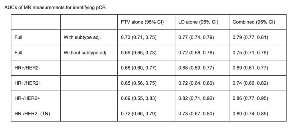
Li W, Onishi N, Wolf DM, et al. MRI models by response predictive subtype for predicting pathologic complete response. 2022 San Antonio Breast Cancer Symposium, 8-11 Dec, 2022; Abstract No. P4-02-10.

Stringer-Reasor E, Shatsky RA, Chien J, et al Evaluation of the PD-1 Inhibitor Cemiplimab in early-stage, high-risk HER2-negative breast cancer: Results from the neoadjuvant I-SPY 2 TRIAL. 2022 San Antonio Breast Cancer Symposium, 8-11 Dec, 2022; Abstract No. PD11-01.

Rosenbluth J, Bui TB, Warhadpande S, et al. Characterization of residual disease after neoadjuvant selective estrogen receptor degrader (SERD) therapy using tumor organoids in the I-SPY Endocrine Optimization Protocol (EOP). 2022 San Antonio Breast Cancer Symposium, 8-11 Dec, 2022; Abstract No. P3-09-01.

Wolf DM, Yau C, Wulfkuhle J, et al. Characterizing the HER2-/Immune-/DNA repair (DRD-) response predictive breast cancer subtype: the hunt for new protein targets in a high-needs population with low response to all I-SPY2 agents. 2022 San Antonio Breast Cancer Symposium, 8-11 Dec, 2022; Abstract No. PD5-04.

Bui TBV, Wolf DM, Moore K,et al. An Organoid Model System to Study Resistance Mechanisms, Predictive Biomarkers, and New Strategies to Overcome Therapeutic Resistance in Early-Stage Triple-Negative Breast Cancer. 2022 San Antonio Breast Cancer Symposium, 8-11 Dec, 2022; Abstract No. PD5-02.

Yee D, Shatsky RA, Yau C, et al. Improved pathologic complete response rates for triple-negative breast cancer in the I-SPY2 Trial. 2022 ASCO Annual Meeting, 3-7 Jun, 2022; Abstract No. 591.

Thomas A, Clark AS, Yau C, et al. Molecular subtype to predict pathologic complete response in HER2-positive breast cancer in the I-SPY2 trial. 2022 ASCO Annual Meeting, 3-7 Jun, 2022; Abstract No. 510.

Huppert LA, Rugo HS, Pusztai L, et al. Pathologic complete response (pCR) rates for HR+/HER2- breast cancer by molecular subtype in the I-SPY2 Trial. 2022 ASCO Annual Meeting, 3-7 Jun, 2022; Abstract No. 504.

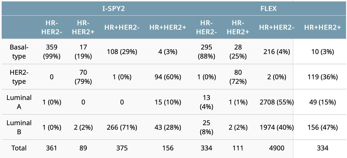
Cha J, Warner P, Hiatt R, et al. Distribution of breast cancer molecular subtypes within receptor classifications: Lessons from the I-SPY2 Trial and FLEX Registry. 2022 ASCO Annual Meeting, 3-7 Jun, 2022; Abstract No. 592.

Mittempergher L, Kuilman MM, Barcaru A, et al. The ImPrint immune signature to identify patients with high-risk early breast cancer who may benefit from PD1 checkpoint inhibition in I-SPY2. 2022 ASCO Annual Meeting, 3-7 Jun, 2022; Abstract No. 514

2021

Kyalwazi B, Yau C, Olopade O, et al. Analysis of clinical outcomes and expression-based immune signatures by race in the I-SPY 2 trial. 2021 San Antonio Breast Cancer Symposium, 7-10 Dec, 2021; Abstract No. GS4-02.

Northrop A, Christofferson A, Melisko M, et al. Improving patient-reported outcome data capture for clinical research: ePRO in ISPY 2, a phase 2 breast cancer study. 2021 San Antonio Breast Cancer Symposium, 7-10 Dec, 2021; Abstract No. P4-12-02.

Goodarzi H, Navickas A, Wang J, et al. Tumor-released circulating orphan non-coding RNAs reflect treatment response and survival in breast cancer.  2021 San Antonio Breast Cancer Symposium, 7-10 Dec, 2021; Abstract No. PD9-04.

Christofferson A, Price E, Mukhtar RA, et al. Single-institution retrospective analysis of lymph node (LN) change on breast MRI in patients with high risk early-stage breast cancer receiving neoadjuvant chemotherapy with and without immunotherapy on the ISPY-2 TRIAL. 2021 San Antonio Breast Cancer Symposium, 7-10 Dec, 2021; Abstract No. P3-03-07.

Bareng TJ, Gibbs JE, Onishi N, et al. Challenges of achieving high image quality on breast MRI for quantitative measurements in the I-SPY 2 TRIAL. 2021 San Antonio Breast Cancer Symposium, 7-10 Dec, 2021; Abstract No. P3-03-04.

Li W, Le NN, Onishi N, et al. Diffusion-weighted MRI for prediction of pathologic complete response in HER2- breast cancer treated with pembrolizumab plus neoadjuvant chemotherapy. 2021 San Antonio Breast Cancer Symposium, 7-10 Dec, 2021; Abstract No. P3-03-02.

Onishi N, Gibbs JE, Li W, et al. Functional tumor volume at 3 and 6-week MRI as an indicator of patients with inferior outcome after neoadjuvant chemotherapy. 2021 San Antonio Breast Cancer Symposium, 7-10 Dec, 2021.
Jones EF, Hathi DK, Konovalova N, et al. Initial experience of FES-dedicated breast PET imaging of early-stage ER+ invasive lobular carcinoma. 2021 San Antonio Breast Cancer Symposium, 7-10 Dec, 2021; Abstract No. P3-02-01.
Jones EF, Hathi DK, Molina-Vega J, et al. FES-dedicated breast PET uptake in early-stage ER+ breast cancers. 2021 San Antonio Breast Cancer Symposium, 7-10 Dec, 2021; Abstract No. P3-02-02.
Chien AJ, Kalinsky KM, Molina-Vega J, et al. I-SPY2 endocrine optimization protocol (EOP): A pilot neoadjuvant endocrine therapy study with amcenestrant as monotherapy or in combination with abemacicilib or letrozole in molecularly selected HR+/HER2- clinical stage 2/3 breast cancer. 2021 San Antonio Breast Cancer Symposium, 7-10 Dec, 2021; Abstract No. OT1-10-02.
Potter DA, Roesch E, Yau C, et al. Evaluation of Tucatinib + (Paclitaxel + Pertuzumab + Trastuzumab) followed by AC in high-risk HER2 positive (HER2+) stage II/III breast cancer: Results from the I-SPY 2 TRIAL. 2021 San Antonio Breast Cancer Symposium, 7-10 Dec, 2021; Abstract No. PD8-07.
Sayaman RW, Wolf DM, Yau C, et al. Elucidating the biology of circulating tumor DNA (ctDNA) shedding across receptor subtypes in high-risk early-stage breast cancer. 2021 San Antonio Breast Cancer Symposium, 7-10 Dec, 2021; Abstract No. P2-01-03.
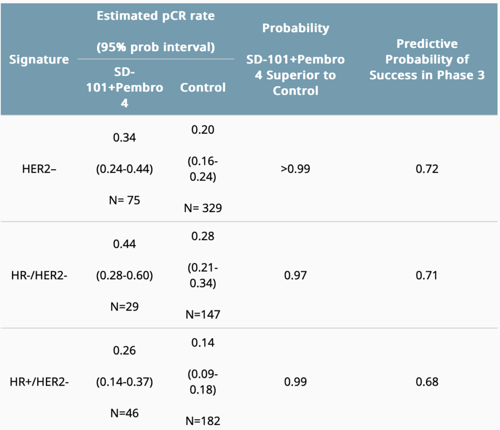
Chien AJ, Soliman HH, Ewing CA, et al. Evaluation of intra-tumoral (IT) SD-101 and pembrolizumab (Pb) in combination with paclitaxel (P) followed by AC in high-risk HER2-negative (HER2-) stage II/III breast cancer: Results from the I-SPY 2 tria. 2021 ASCO Annual Meeting, 4-8 Jun, 2021; Abstract No. 508.
Marczyk M, Mrukwa A, Yau C, et al. Treatment Efficacy Score (TES), a continuous residual cancer burden-based metric to compare neoadjuvant chemotherapy efficacy between trial arms in the I-SPY 2 trial. 2021 ASCO Annual Meeting, 4-8 Jun, 2021; Abstract No. 587.
Soliman H, Wolf D, Chien J, et al. Chemokine12 (CK12) tertiary lymphoid gene expression signature as a predictor of response in 3 immunotherapy arms of the neoadjuvant ISPY 2 TRIAL – pembrolizumab with and without SD101, and durvalumab combined with olaparib – and in 9 other arms of the trial including platinum- based and dual-anti-HER2 therapies. 2021 San Antonio Breast Cancer Symposium, 7-10 Dec, 2021; Abstract No. PD10-07.

2020

Yee D, Haluska P, Wolf DM, et al. Biomarker analysis of paclitaxel, ganitumab, and metformin (PGM) therapy in the I-SPY2 neoadjuvant clinical trial. 2020 San Antonio Breast Cancer Symposium, Dec 8-11, 2020; Abstract No. PS4-08.

Wolf DM, Yau C, Brown-Swigart L, et al. Biomarkers predicting response to durvalumab combined with olaparib in the neoadjuvant I-SPY 2 TRIAL for high-risk breast cancer. 2020 San Antonio Breast Cancer Symposium, Dec 8-11, 2020; Abstract No. PD14-02.

Shad S, van der Noordaa M, Osdoit M, et al. Site of recurrence after neoadjuvant therapy: a multi-center pooled analysis. 2020 San Antonio Breast Cancer Symposium, Dec 8-11, 2020; Abstract No. PD13-02.

van der Noordaa MEM, Yau C, Shad S, et al. Assessing prognosis after neoadjuvant therapy: A comparison between anatomic ypAJCC staging, Residual Cancer Burden Class and Neo-Bioscore. 2020 San Antonio Breast Cancer Symposium, Dec 8-11, 2020; Abstract No. GS4-07.

Li W, Newitt DC, Gibbs J, et al. Subtype-specific MRI models to guide selection of candidates for de-escalation of neoadjuvant therapy. 2020 San Antonio Breast Cancer Symposium, Dec 8-11, 2020; Abstract No. PD6-05.

Magbanua MJM, Wolf D, Renner D, et al. Personalized circulating tumor DNA (ctDNA) as predictive biomarker in high-risk early stage breast cancer (EBC) treated with neoadjuvant chemotherapy (NAC) with or without pembrolizumab (P). 2020 San Antonio Breast Cancer Symposium, Dec 8-11, 2020; Abstract No. PD9-02.

Wulfkuhle J, Wolf D, Yau C, et al. Identification of biomarkers associated with therapeutic resistance: quantitative protein/phosphoprotein analysis of ~750 patients across 8 arms of the neoadjuvant I-SPY 2 TRIAL for high-risk early stage breast cancer. 2020 San Antonio Breast Cancer Symposium, Dec 8-11, 2020; Abstract No. PD9-04.
Yu K, Basu A, Yau C, et al. Computational drug repositioning for the identification of new agents to sensitize drug-resistant breast tumors across treatment arms and molecular subtypes. 2020 San Antonio Breast Cancer Symposium, Dec 8-11, 2020; Abstract No. PS11-04.
Gibbs J, Newitt DC, Watkins M, et al. Operational standardization and quality assurance yield high acceptance rate for breast MRI in the I-SPY 2 TRIAL. 2020 San Antonio Breast Cancer Symposium, Dec 8-11, 2020; Abstract No. PS11-08.
Beckwith H, Schwab R, Yau C, et al. Evaluation of SGN-LIV1a followed by AC in high-risk HER2 negative stage II/III breast cancer: Results from the I-SPY 2 TRIAL. 2020 San Antonio Breast Cancer Symposium, Dec 8-11, 2020; Abstract No. PD1-10.
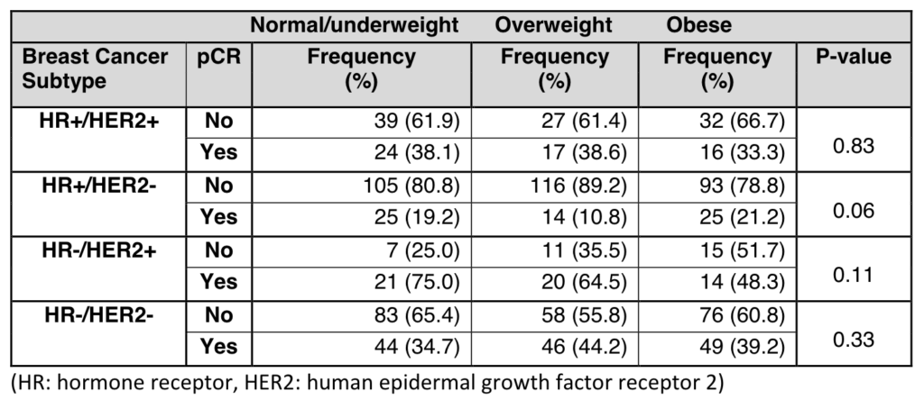
Wang H, Yee D, Potter D, et al. Impact of Body Mass Index on Pathological Complete Response after Neoadjuvant Chemotherapy: Results from the I-SPY 2 trial. 2020 San Antonio Breast Cancer Symposium, Dec 8-11, 2020; Abstract No. PS6-05.
Onishi N, Li W, Venters SJ, et al. Radiologic review to refine selection of candidates for de-escalation of neoadjuvant therapy after mid-treatment biopsy. 2020 San Antonio Breast Cancer Symposium, Dec 8-11, 2020; Abstract No. PS3-02.
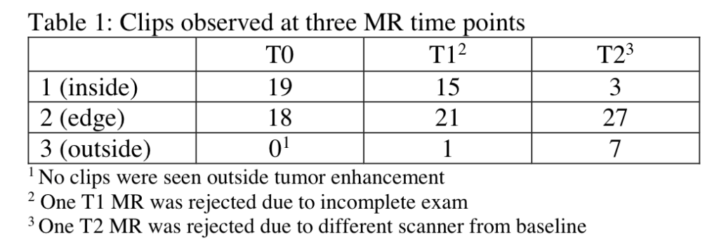
Bareng TJ, Gibbs J, Li W, et al. Assessment of clip locations on breast MRI during NAC to guide tumor bed biopsy at mid-treatment. 2020 San Antonio Breast Cancer Symposium, Dec 8-11, 2020; Abstract No. PS3-14.
Hathi DK, Jones EF, Li W, et al. Relationship of dedicated breast PET and MRI features in breast cancer patients receiving neoadjuvant chemotherapy. 2020 San Antonio Breast Cancer Symposium, Dec 8-11, 2020; Abstract No. PS13-50.
Venters SJ, Li W, Wolf DM, et al. Serial MRI and pathology combined to select candidates for therapy de-escalation in the I-SPY 2 TRIAL. 2020 San Antonio Breast Cancer Symposium, Dec 8-11, 2020; Abstract No. PS4-10.
Carter JM, Klein ME, Venters SJ, et al. Pathologic features of the inter-regimen biopsy predict response to neoadjuvant therapy in the I-SPY2 trial. 2020 San Antonio Breast Cancer Symposium, Dec 8-11, 2020; Abstract No. PS4-09.
Magbanua MJM, van ‘t Veer L, Clark A, et al. Outcomes associated with disseminated tumor cells at surgery after neoadjuvant chemotherapy in high-risk early stage breast cancer: the I-SPY SURMOUNT Study. 2020 San Antonio Breast Cancer Symposium, Dec 8-11, 2020; Abstract No. PS2-07.
Pusztai L, Han HS, Yau C, et al. Evaluation of durvalumab in combination with olaparib and paclitaxel in high-risk HER2 negative stage II/III breast cancer: Results from the I-SPY 2 TRIAL. 2020 AACR, June 20-24, 2020; Abstract No. CT011.

2019

Pohlmann PR, Yau C, DeMichele A, et al. Amsterdam 70-gene profile (MammaPrint) Low Risk, even in the HER2 positive subset, identifies a population of women with lower early risk for recurrence despite low response rates to chemotherapy and to HER2 targeted therapy. 2019 San Antonio Breast Cancer Symposium, December 10-14, 2019; Abstract No. P6-10-06.

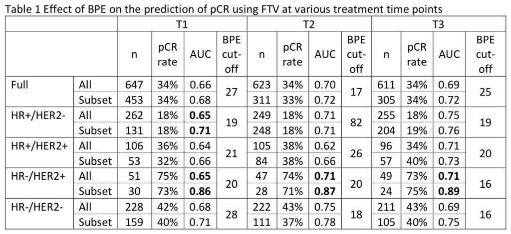
Li W, Onishi N, Newitt DC, et al. The effect of background parenchymal enhancement on the predictive performance of functional tumor volume measured in MRI. 2019 San Antonio Breast Cancer Symposium, December 10-14, 2019; Abstract No. P6-02-01.

Venters SJ, Wolf DM, Brown-Swigart L, et al. Assessing biomarkers to inform treatment de-escalation: mid-treatment biopsy cellularity predicts pCR in the I-SPY 2 TRIAL. 2019 San Antonio Breast Cancer Symposium, December 10-14, 2019; Abstract No. P6-10-02.

Magbanua MM, Brown-Swigart L, Hirst G, et al. Personalized monitoring of circulating tumor DNA during neoadjuvant therapy in highrisk early stage breast cancer reflects response and risk of metastatic recurrence. 2019 San Antonio Breast Cancer Symposium, December 10-14, 2019; Abstract No. P5-01-04.

Ghersin H, Chattopadhyay A, Blaes A, et al. Introducing an electronic platform to collect patient reported outcomes in the I-SPY 2 TRIAL, a neoadjuvant clinical trial. 2019 San Antonio Breast Cancer Symposium, December 10-14, 2019; Abstract No. OT3-19-02.

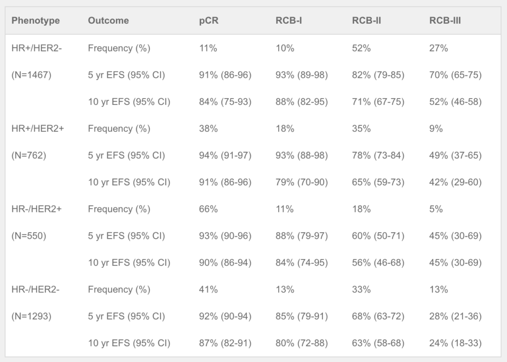
Yau C, van der Noordaa M, Wei J, et al. Residual cancer burden after neoadjuvant therapy and long-term survival outcomes in breast cancer: a multi-center pooled analysis. 2019 San Antonio Breast Cancer Symposium, December 10-14, 2019; Abstract No. GS5-01.

Wolf DM, Yau C, Wulfkuhle J, et al. HER2 signaling, ER, and proliferation biomarkers predict response to multiple HER2-targeted agents/combinations plus standard neoadjuvant therapy in the I-SPY 2 TRIAL. 2019 San Antonio Breast Cancer Symposium, December 10-14, 2019; Abstract No. P4-10-02.
Sayaman RW, Wolf DM, Yau C, et al. Application of Machine Learning to elucidate the biology predicting response in the I‑SPY 2 neoadjuvant breast cancer trial. 2019 San Antonio Breast Cancer Symposium, December 10-14, 2019; Abstract No. P1-21-08.
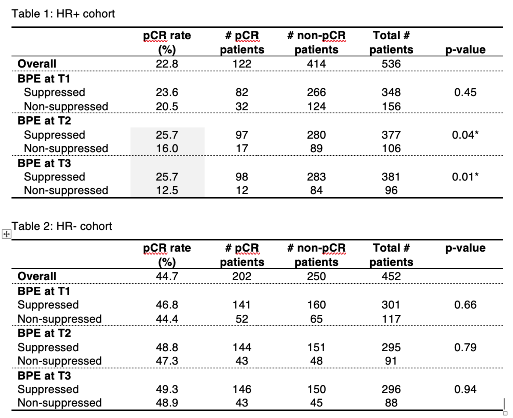
Onishi N, Li W, Newitt DC, et al. Lack of background parenchymal enhancement suppression in breast MRI during neoadjuvant chemotherapy may be associated with inferior treatment response in hormone receptor positive breast cancer. 2019 San Antonio Breast Cancer Symposium, December 10-14, 2019; Abstract No. PD9-05.
Li W, Onishi N, Newitt DC, et al. Breast cancer subtype specific association of pCR with MRI assessed tumor volume progression during NAC in the I-SPY 2 TRIAL. 2019 San Antonio Breast Cancer Symposium, December 10-14, 2019; Abstract No. PD9-04.
Arasu V, Kim P, Li W, et al. Additive benefit of MRI background parenchymal enhancement (BPE) to an MRI tumor volume model for predicting pathologic complete response (pCR) to neoadjuvant treatment in I-SPY2. 2019 San Antonio Breast Cancer Symposium, December 10-14, 2019; Abstract No. PD9-06.
Osdoit M, Yau C, Symmans WF, et al. The impact of residual ductal carcinoma in situ on breast cancer recurrence in the neoadjuvant I-SPY2 TRIAL. 2019 San Antonio Breast Cancer Symposium, December 10-14, 2019; Abstract No. P3-08-16.
Helsten TL, Lo SS, Yau C, et al. Evaluation of patritumab/paclitaxel/trastuzumab over standard paclitaxel/trastuzumab in early stage, high-risk HER2 positive breast cancer: Results from the neoadjuvant I-SPY 2 TRIAL. 2019 San Antonio Breast Cancer Symposium, December 10-14, 2019; Abstract No. P3-11-02.
Liu MC, Robinson PA, Yau C, et al. Evaluation of a pembrolizumab-8 cycle neoadjuvant regimen without AC for high-risk early-stage HER2-negative breast cancer: Results from the I-SPY 2 TRIAL. 2019 San Antonio Breast Cancer Symposium, December 10-14, 2019; Abstract No. P3-09-02.
Basu A, Philip EJ, Dewitt B, et al. The Clinical Benefit Index: A Pilot Study Integrating Treatment Efficacy and Quality of Life in Oncology Clinical Trials. 2019 San Antonio Breast Cancer Symposium, December 10-14, 2019; Abstract No. P2-12-06.
Yau C, Symmans W, Pusztai L, et al. Site of recurrence after neoadjuvant therapy: clues to biology and impact on endpoints. 2019 San Antonio Breast Cancer Symposium, December 10-14, 2019; Abstract No. P2-20-02.
Du L, Yau C, Brown-Swigart L, et al. Prognostic contribution of predicted sensitivity to endocrine therapy (SET) prior to neoadjuvant chemotherapy for stage II-III hormone receptor-positive and HER2- negative (HR+/HER2-) breast cancer. 2019 San Antonio Breast Cancer Symposium, December 10-14, 2019; Abstract No. P2-11-04.
Campbell MJ, Yau C, Bolen J, et al. Analysis of immune cell infiltrates as predictors of response to the checkpoint inhibitor pembrolizumab in the neoadjuvant I-SPY 2 TRIAL. AACR Annual Meeting, Mar 29 – Apr 3, 2019. In: Cancer Res 2019; 79 (13 Suppl); Abstract No. CT003.
Li W, Newitt DC, Wilmes LJ, et al. Combination of MRI quantitative measures improves prediction of residual disease following neoadjuvant chemotherapy (NAC) for breast cancer in the I-SPY 2 TRIAL. ISMRM 27th Annual Meeting, May 11-16, 2019; Abstract No. 0278.
Wulfkuhle JD, Yau C, Wolf D, et al. Quantitative MHC II protein expression levels in tumor epithelium to predict response to the PD1 inhibitor pembrolizumab in the I-SPY 2 Trial. 2019 ASCO Annual Meeting, May 30 – June 4, 2019. In: J Clin Oncol 2019; 37 (15); Abstract No. 2631.
Yau C, van der Noordaa M, Wei J, et al. Residual cancer burden after neoadjuvant therapy and long-term survival outcomes in breast cancer: A multi-center pooled analysis. 2019 San Antonio Breast Cancer Symposium, December 10-14, 2019. In: Cancer Research 2020; 80 (4_Supplement); Abstract No. GS5-01.

2018

Hyland C, Varghese F, Yau C, et al. The use of 18F-FDG PET/CT as an initial staging procedure for stage II-III breast cancer reduces false positives, costs, and time to treatment: a multicenter value analysis in the I-SPY2 trial. 2018 San Antonio Breast Cancer Symposium, December 4-8, 2018; Abstract No. P5-15-01.

Zhu Z, Yau C, van ’t Veer L, et al. Molecular subtypes of invasive lobular breast cancer in the I-SPY2 Trial. 2018 San Antonio Breast Cancer Symposium, December 4-8, 2018; Abstract No. PD7-06.
Yau C, Wolf D, Campbell M, et al. Expression-based immune signatures as predictors of neoadjuvant targeted-/chemo-therapy response: Experience from the I-SPY 2 TRIAL of ~1000 patients across 10 therapies. 2018 San Antonio Breast Cancer Symposium, December 4-8, 2018; Abstract No. P3-10-06.
Yau C, Brown-Swigart L, Asare S, et al. LIV-1 Expression in Primary Breast Cancers in the I-SPY 2 TRIAL. 2018 San Antonio Breast Cancer Symposium, December 4-8, 2018; Abstract No. P3-10-14.
Li W, Newitt DC, Yun BL, et al. MRI detection of residual disease following neoadjuvant chemotherapy (NAC) in the I-SPY 2 TRIAL. 2018 San Antonio Breast Cancer Symposium, December 4-8, 2018; Abstract No. PD4-03.
Hylton NM, Symmans WF, Yau C, et al. Refining neoadjuvant predictors of three year distant metastasis free survival: integrating volume change as measured by MRI with residual cancer burden. 2018 San Antonio Breast Cancer Symposium, December 4-8, 2018; Abstract No. P2-07-03.
van der Noordaa MEM, Esserman L, Yau C, et al. Role of breast MRI in predicting pathologically negative nodes after neoadjuvant chemotherapy in cN0 patients in the I-SPY 2 trial. 2018 San Antonio Breast Cancer Symposium, December 4-9, 2018; Abstract No. PD4-04.
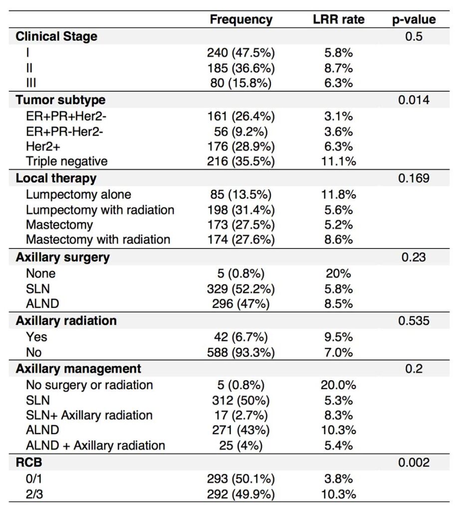
Silverstein J, Suleiman L, Yau C, et al. The impact of local therapy on locoregional recurrence in women with high risk breast cancer in the neoadjuvant I-SPY2 TRIAL. 2018 San Antonio Breast Cancer Symposium, December 4-8, 2018; Abstract No. P2-14-01.
Wolf DM, Yau C, Wulfkhule J, et al. Identifying breast cancer molecular phenotypes to predict response in a modern treatment landscape: lessons from ~1000 patients across 10 arms of the I-SPY 2 TRIAL. 2018 San Antonio Breast Cancer Symposium, December 4-8, 2018; Abstract No. P3-10-02.
Magbanua M, Brown-Swigart L, Hirst G, et al. Personalized serial circulating tumor DNA (ctDNA) analysis in high-risk early stage breast cancer patients to monitor and predict response to neoadjuvant therapy and outcome in the I-SPY 2 TRIAL. 2018 San Antonio Breast Cancer Symposium, Dec 4-8, 2018; Abstract No. PD2-01.
van ‘t Veer L, Wolf D, Yau C, et al. MammaPrint High1/High2 risk class as a pre-specified biomarker of response to nine different targeted agents plus standard neoadjuvant therapy for ~ 1000 breast cancer patients in the I-SPY 2 TRIAL. 30th EORTC-NCI-AACR Symposium, November 13-16, 2018
Gallagher RI, Wulfkuhle JD, Yau C, et al. Association of activation levels of TIE2 with response to the angiogenesis inhibitor trebananib in HER2+ patients in the I-SPY 2 trial. 2018 American Society of Cancer Oncology Annual Meeting, June 1-5, 2018. In: J Clin Oncol 2018; 36 (suppl); Abstract No. 12103.
Wulfkuhle JD, Wolf DM, Yau C, et al. Phosphorylation of AKT kinase substrates to predict response to the AKT inhibitor MK2206 in the I-SPY 2 trial in both HER2- and HER2+ patients. 2018 American Society of Cancer Oncology Annual Meeting, June 1-5, 2018. In: J Clin Oncol 2018; 36 (suppl); Abstract No. 12099.
Fraser Symmans W, Yau C, Chen Y, et al. Residual cancer burden (RCB) as prognostic in the I-SPY 2 TRIAL. 2018 American Society of Cancer Oncology Annual Meeting, June 1-5, 2018. In: J Clin Oncol 2018; 36 suppl; Abstract No. 520.
William Fraser Symmans, Christina Yau, Yunn-Yi Chen, et al. Advances in Precision Medicine in Triple-Negative Breast Cancer: Residual cancer burden (RCB) as prognostic in the I-SPY 2 TRIAL Poster Presentation. 2018 American Society of Cancer Oncology Annual Meeting, June 1-5, 2018. In: J Clin Oncol 2018; 36 (suppl); Abstract No. 520.
Wolf DM, Yau C, Brown-Swigart L, et al. Evaluation of ANG/TIE/hypoxia pathway genes and signatures as predictors of response to trebananib (AMG 86) in the neoadjuvant I-SPY 2 TRIAL for Stage II-III high-risk breast cancer. 2018 AACR Annual Meeting, April 14-18, 2018. In: Cancer Res 2018; 78 (13 Suppl); Abstract No. 2611.
Lee PRE, Zhu Z, Wolf D, et al. BluePrint Luminal subtype predicts non-response to HER2-targeted therapies in HR+/HER2+ I-SPY 2 breast cancer patients. 2018 AACR Annual Meeting, April 14-18, 2018. In: Cancer Res 2018; 78 (13 Suppl); Abstract No. 2612.

2017

Campbell M, Yau C, Borowsky A, et al. Analysis of immune infiltrates (assessed via multiplex fluorescence immunohistochemistry) and immune gene expression signatures as predictors of response to the checkpoint inhibitor pembrolizumab in the neoadjuvant I-SPY 2 trial. 2017 San Antonio Breast Cancer Symposium, Dec 5-9, 2017. In: Cancer Res 2018; 78 (4 Suppl); Abstract No. PD6-08.
Yee D, DeMichele A, Isaacs C, et al. Pathological complete response predicts event-free and distant disease-free survival in the I-SPY2 TRIAL. 2017 San Antonio Breast Cancer Symposium, Dec 5-9, 2017. In: Cancer Res 2018; 78 (4 Suppl); Abstract No. GS3-08.
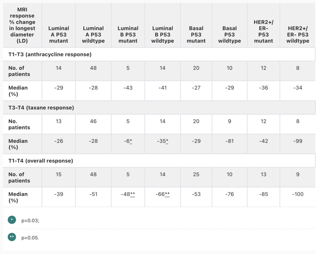
Pradhan SM, Carey L, Edmiston S, et al. P53 mutation and differential response to neoadjuvant chemotherapy in women with locally advanced breast cancer: Results from the I-SPY trial (CALGB 150007/1500012 and ACRIN 6657). 2017 American Society of Clinical Oncology Annual Meeting, June 2-6, 2017. In: J Clin Oncol 2009; 27 (15 Suppl.); Abstract No. 11099.
Nanda R, Liu MC, Yau C, et al. Pembrolizumab plus standard neoadjuvant therapy for high-risk breast cancer (BC): Results from I-SPY 2. 2017 American Society of Clinical Oncology Annual Meeting, June 2-6, 2017. In: J Clin Oncol 2017; 35 (15 Suppl.); Abstract No. 506.
Partridge SC, Zhang Z, Newitt DC, et al. ACRIN 6698 trial: Quantitative diffusion-weighted MRI to predict pathologic response in neoadjuvant chemotherapy treatment of breast cancer. 2017 American Society of Clinical Oncology Annual Meeting, June 2-6, 2017. In: J Clin Oncol 2017; 35 (15 Suppl.); Abstract No. 11520.
Li W, Wilmes LJ, Newitt DC, et al. Diffusion-weighted MRI improves imaging prediction of response in the I-SPY 2 trial. 2017 San Antonio Breast Cancer Symposium, December 5-9, 2017. In: Cancer Res 2018; 78 (4 Suppl.); Abstract No. P2-09-23.
Vidula N, Yau C, Rugo HS, et al. Trop2 gene expression (Trop2e) in primary breast cancer (BC): Correlations with clinical and tumor characteristics. 2017 American Society of Cancer Oncology Annual Meeting, June 2-6, 2017. In: J Clin Oncol 2017; 35 (15 Suppl.); Abstract No. 1075.
Wolf D, Yau C, Brown-Swigart L, et al. Analysis of biomarkers for response and resistance to the AKT inhibitor MK-2206 in the neoadjuvant I-SPY 2 trial for stage II-III high-risk breast cancer. 2017 San Antonio Breast Cancer Symposium, December 5-9, 2017. In: Cancer Res 2018; 78 (4 Suppl.); Abstract No. P2-09-08.
Yau C, Wolf D, Brown-Swigart L, et al. Analysis of DNA repair deficiency biomarkers as predictors of response to the PD1 inhibitor pembrolizumab: Results from the neoadjuvant I-SPY 2 trial for stage II-III high-risk breast cancer. 2017 San Antonio Breast Cancer Symposium, December 5-9, 2017. In: Cancer Res 2018; 78 (4 Suppl.); Abstract No. PD6-14.

2016

Vidula N, Yau C, Wolf DM, et al. Androgen receptor (AR) expression in primary breast cancer (BC): Correlations with clinical characteristics and outcomes. 2016 American Society of Clinical Oncology Annual Meeting, June 3-7, 2016. In: J Clin Oncol 2016; 34 (15 Suppl.), Abstract No. 1072.
DeMichele AM, Moulder S, Buxton M, et al. Efficacy of T-DM1 + pertuzumab over standard therapy for HER2+ breast cancer: Results from the neoadjuvant I-SPY 2 TRIAL. AACR 107th Annual Meeting 2016, April 16-20, 2016. In: Cancer Res 2016; 76 (14 Suppl.), Abstract No. CT042.
Buxton M, DeMichele AM, Chia S, et al. Efficacy of pertuzumab/trastuzumab/paclitaxel over standard trastuzumab/paclitaxel therapy for HER2+ breast cancer: Results from the neoadjuvant I-SPY 2 TRIAL. AACR 107th Annual Meeting 2016, April 16-20, 2016. In: Cancer Res 2016; 76 (14 Suppl.), Abstract No. CT106.
Li W, Arasu V, Jones EF, et al. Effect of MR imaging contrast kinetic thresholds for prediction of neoadjuvant chemotherapy response in breast cancer subtypes – Results from ACRIN 6657 / I-SPY 1 trial. 2016 San Antonio Breast Cancer Symposium, December 6-10, 2016. In: Cancer Res 2016; 77 (4 Suppl.), Abstract No. PD3-05.
Wolf DM, Yau C, Brown-Swigart L, et al. Gene and pathway differences between MammaPrint High1/High2 risk classes: results from the I-SPY 2 TRIAL in breast cancer. AACR 107th Annual Meeting 2016, April 16-20, 2016. In: Cancer Res 2016; 76 (14 Suppl.), Abstract No. 859.
Wolf DM, Yau C, Sanil A, et al. Combining sensitivity markers to identify triple-negative breast cancer patients most responsive to veliparib/carboplatin: results from the I-SPY 2 TRIAL. AACR 107th Annual Meeting 2016, April 16-20, 2016. In: Cancer Res 2016; 76 (14 Suppl.), Abstract No. 858.
Yee D, Paoloni M, Van’t Veer L, et al. The evaluation of ganitumab/metformin plus standard neoadjuvant therapy in high-risk breast cancer: Results from the I-SPY 2 trial. 2016 San Antonio Breast Cancer Symposium, December 6-10, 2016. In: Cancer Res 2016; 77 (4 Suppl.), Abstract No. P6-11-04.
Shah M, Jensen R, Yau C, et al. Trajectory of patient (Pt) reported physical function (PF) during and after neoadjuvant chemotherapy in the I-SPY 2 trial. 2016 San Antonio Breast Cancer Symposium, December 6-10, 2016. In: Cancer Res 2017; 77 (4 Suppl.), Abstract No. P5-11-18.
Paoloni M, Lyandres J, Buxton MB, et al. A longitudinal look at toxicity management within a platform trial: Lessons from the I-SPY 2 TRIAL. 2016 San Antonio Breast Cancer Symposium, December 6-10, 2016. In: Cancer Res 2016; 77 (4 Suppl.), Abstract No. P2-11-02.
Forero A, Yee D, Buxton MB, et al. Efficacy of Hsp90 inhibitor ganetespib plus standard neoadjuvant therapy in high-risk breast cancer: Results from the I-SPY 2 trial. 2016 San Antonio Breast Cancer Symposium, December 6-10, 2016. In: Cancer Res 2016; 77 (4 Suppl.), Abstract No. P6-11-02.
Gallagher RI, Yau C, Wolf DM, et al. Quantitative ERα measurements in TNBC from the I-SPY 2 TRIAL correlate with HER2-EGFR co-activation and heterodimerization. 2016 San Antonio Breast Cancer Symposium, December 6-10, 2016. In: Cancer Res 2016; 77 (4 Suppl.), Abstract No. P3-05-02.
Wolf DM, Yau C, Sanil A, et al. DNA repair deficiency biomarkers and MammaPrint high1/(ultra)high2 risk as predictors of veliparib/carboplatin response: Results from the neoadjuvant I-SPY 2 trial for high risk breast cancer. 2016 San Antonio Breast Cancer Symposium, December 6-10, 2016. In: Cancer Res 2016; 77 (4 Suppl.), Abstract No. S2-06.

Abstract No. GS5-04
2022 San Antonio Breast Cancer Symposium, 8-11 Dec, 2022

Identification of symptoms that are associated with irAEs in the I-SPY clinical trial

Basu A, Umashankar S, Melisko M, Lu R, Yu H, Yau C, Asare S, Pitsouni M, Shatsky RA, Isaacs C, DeMichele A, Hershman D, Nanda R, Kim MO, Esserman LJ, Rugo H

Background. Immunotherapy has emerged as an important component of neoadjuvant therapy for some patients with breast cancer (BC). As a result, immune-related adverse events (irAEs) are increasing and have effects on both short and long term symptoms significantly impacting patient quality of life. BC patients may develop new conditions including arthralgias, gastrointestinal issues, endocrinopathies, and fatigue during or after cancer therapy that may be acute or long-lasting in nature. Monitoring for early onset and severity of symptoms, and adjusting treatment and symptom management could optimize therapy for a particular patient, maximizing potential efficacy while mitigating toxicity. We sought to identify patient demographic characteristics and symptom patterns associated with risk for development of irAEs in the context of a randomized trial for patients with early-stage high-risk breast cancer.

Methods. I-SPY2 is a multi-center, phase 2 trial using response-adaptive randomization for high-risk early-stage women with BC. The study population for this analysis includes enrolled patients receiving combinations of experimental immunotherapy and chemotherapy. Groups considered for statistical comparisons included those that developed an irAE versus those that did not develop an irAE up until the surgery timepoint. In I-SPY adverse events are documented through the Common Terminology Criteria for Adverse Events (CTCAEv5.0). Hypothyroidism, adrenal insufficiency, and pneumonitis were the irAEs considered in this study. A chi-square test was used to assess associations between race and ethnicity (White, Asian, Black, non-Hispanic) and irAEs. One-way ANOVA was used to evaluate the association between age (>50 vs < 50) and irAEs. 33 symptoms reported at CTCAE grade 2 or higher were included in the analyses and a symptom burden score was calculated using area under curve (AUC) which combined the duration of each symptom between baseline and week 6 of treatment, and grade of adverse event. Regularized regression using leave-one out cross validation was used to evaluate early symptoms (as quantified by the symptom burden score) as predictors, and irAEs as surrogate responses.

Results. Out of 461 patients, percentages of patients wth irAEs of interest included hypothyroidism (13%), adrenal insufficiency (9%), and pneumonitis (4%). Demographic information was available for 333 patients, of which 270 (81%) were White, 23 (7%) were Asian, 37 (11%) were African American (AA) and 278 (17%) were non-Hispanic. There were proportionately higher number of white patients that developed hypothyroidism than non-white patients (35 of 265 (13%) vs 2 of 63 (3%), P < 0.04). Pneumonitis was more common in patients over 50 years old than under 50 years old (P < 0.02). Symptoms that were most commonly reported up to week 6 of treatment among patients who developed an irAE included: diarrhea (36%), fatigue (15%), dizziness (12%) and shortness of breath (SOB) (11%). Symptoms associated with the development of hypothyroidism included fatigue (15%, mean AUC=11.8 vs 5.8 for those that did not develop irAE), SOB (11%, 4.3 vs 2.8), and blurry vision (1%, 1.0 vs 0.12). Development of adrenal insufficiency was associated with early reports of diarrhea (36%, 19.0 vs 10.5), SOB (11%, 7.8 vs 2.6), joint pain (3%, 2.29 vs 0.58), decreased appetite (3%, 3.55 vs 0.91), and constipation (1%, 3.6 vs 0.02). No significant early symptoms emerged for pneumonitis due to a limited number of events.

Conclusion. Our study utilizes an analysis framework that is aimed to determine symptom clusters that predict the development of irAEs. We describe specific symptoms presenting early with the development of hypothyroidism and adrenal insufficiency, in recognition of allowing physicians to be more diligent in active and post treatment monitoring.

Abstract No. GS5-03
2022 San Antonio Breast Cancer Symposium, 8-11 Dec, 2022

Evaluation of anti-PD-1 Cemiplimab plus anti-LAG-3 REGN3767 in early-stage, high-risk HER2-negative breast cancer: Results from the neoadjuvant I-SPY 2 TRIAL

Isaacs C, Nanda R, Chien J, Trivedi MS, Stringer-Reasor E, Vaklavas C, Boughey JC, Sanford A, Wallace A, Clark AS, Thomas A, Albain KS, Kennedy LC, Sanftt TB, Kalinsky K, Han HS, Williams N, Arora M, Elias A, Falkson C, Asare S, Lu R, Pitsouni M, Wilson A, Perlmutter J, Rugo H, Schwab R, Symmans WF, Hylton NM, van ‘t Veer L, Yee D, DeMichele A, Berry D, Esserman LJ, I-SPY Investigators

Background: I-SPY2 is a multicenter, phase 2 trial using response-adaptive randomization within biomarker subtypes defined by hormone-receptor (HR), HER2, and MammaPrint (MP) status to evaluate novel agents as neoadjuvant therapy for high-risk breast cancer. The primary endpoint is pathologic complete response (pCR). Cemiplimab is an anti-PD-1 inhibitor approved for the treatment of NSCLC and cutaneous basal and squamous cell CA. Lymphocyte activation gene 3 (LAG-3) binds MHC class II leading to inhibition of T-cell proliferation and activation and is often co-expressed with PD-1. REGN3767 is a fully humanized mAb that binds to LAG-3 and blocks inhibitory T-cell signaling. Concurrent blockade of LAG-3 with an anti-PD-1 may enhance efficacy of an anti-PD-1.  

Methods: Women with tumors ≥ 2.5cm were eligible for screening. Only HER2 negative (HER2-) patients were eligible for this treatment; HR positive (HR+) patients had to be MP high risk. Treatment included Paclitaxel 80 mg/m2 IV weekly x 12 and Cemiplimab 350 mg and REGN3767 1600 mg both given q3weeks x 4, followed by doxorubicin/cyclophosphamide (AC) every 2 weeks x 4. The control arm was weekly paclitaxel x 12 followed by AC every 2-3 weeks x 4. Cemiplimab/REGN3767 was eligible to graduate in 3 of 10 pre-defined signatures: HER2-, HR-HER2-, and HR+HER2-. The statistical methods for evaluating I-SPY 2 agents has been previously described. To adapt to changing standard of care, we constructed “dynamic controls” comprising ‘best’ alternative therapies using I-SPY 2 and external data and estimated the probability of Cemiplimab/REGN3767 being superior to the dynamic control. Response predictive subtypes (Immune+ vs Immune-) were assessed using pre-treatment gene expression data and the ImPrint signature.  

Results: 73 HER2- patients (40 HR+ and 33 HR-) received Cemiplimab/REGN3767 treatment. The control group included [357 patients with HER2- tumors (201 HR+ and 156 HR-) enrolled since March 2010. Cemiplimab/REGN3767 graduated in both HR-/HER2- and HR+/HER2- groups; estimated pCR rates (as of June 2022) are summarized in the table. Safety events of note for Cemiplimab/REGN3767 include hypothyroidism 30.8%, adrenal insufficiency (AI) 19.2%, hyperthyroidism 14.1%, pneumonitis 1.3%, and hepatitis 3.8%. All were G1/2 except for 6 (7.7%) G3 AI and 3 (3.8%) G3 colitis. Rash occurred in 62.8%, 9% G3 and 2 pts (2.6%) had pulmonary embolism. X% of adrenal insufficiency cases required replacement therapy. 40 patients (11 HR+ and 29 HR-) in Cemiplimab/REGN3767 were predicted Immune+; 32 (29 HR+ and 3 HR-) were predicted Immune-. In the HR+ group pCR was achieved in 10/11 (91%) patients with Immune+ subtype compared with 8/29 (28%) with Immune- subtype. Additional biomarker analyses are ongoing and will be presented at the meeting.

Conclusion: The I-SPY 2 study aims to assess the probability that investigational regimens will be successful in a phase 3 neoadjuvant trial. Dual immune blockade with a LAG-3 inhibitor and anti-PD1 therapy resulted in a high predicted pCR rate both in HR-/HER2- (60%) and HR+/HER2- (37%) disease. The novel Imprint signature identified a group of HR+ patients most likely to benefit from this active regimen.

Abstract No. P6-01-33
2022 San Antonio Breast Cancer Symposium, 8-11 Dec, 2022

Diffusion-weighted magnetic resonance imaging for subtype-specific prediction of pathologic complete response in neoadjuvant chemotherapy

Zimmerman J, Carmona-Bozo J, Le NN, Onishi N, Wilmes LJ, Gibbs JE, Liang J, Newitt DC, Partridge S, Bolan P, Joe BN, Price ER, LeStage B, Hylton NM, Li W

Background: The apparent diffusion coefficient (ADC) presents a biomarker that is sensitive to tumor cellularity. ADC maps can be calculated from non-contrast diffusion-weighted magnetic resonance imaging (DW-MRI) measurements. ACRIN 6698, a sub-study of clinical trial I-SPY 2, investigated mean ADC – averaged over the whole tumor – as a marker to predict pathologic complete response (pCR) [1]. This work compares a group of histogram-based ADC metrics in addition to mean ADC for early prediction of pCR in patients stratified by breast cancer subtype.

Methods: We performed a retrospective analysis of DW-MRI, dynamic-contrast enhanced (DCE) MRI, and clinical outcome (i.e., pCR at surgery) in a cohort of 79 female patients who were diagnosed with high-risk, stage II/III breast cancer. Patients underwent neoadjuvant chemotherapy (NAC) with paclitaxel (12 weeks), followed by doxorubicin plus cyclophosphamide (12 weeks). The included population represents a subset of the I-SPY 2 cohort and comprises 48 patients with hormone receptor [HR]+/HER2-, and 31 patients with HR-/HER2-. DW- and DCE-MRI acquisitions were performed according to the I-SPY 2 protocol at pretreatment (T0) and after three weeks (T1) and were analyzed to find early treatment percentage (%) change (T0 to T1) in any metric M; where %-change = 100 × (M(T1) – M(T0))/M(T0). Histogram analysis provided nine region-of-interest (ROI)-based ADC metrics (Table 1). ROIs were manually delineated by expert observers in three-dimensional ADC maps, focusing on diffusion-restricted regions [2]. DCE-MRI was analyzed for the integral I-SPY 2 imaging marker of %-change in functional tumor volume (FTV) between T0 and T1. Statistical analysis compared the predictive power of ADC metrics and FTV, including: the receiver-operating-characteristic (ROC) curve from a logistic regression model to predict pCR as ‘positive’, area-under-the-curve (AUC) assessment, and rank-sum Wilcoxon test (p < 0.05: statistically significant).

Results: (Table 1): 16 out of 79 (20.3%) patients reached pCR at surgery, with 18.8% pCR among HR+/HER- and 22.6% among HR-/HER2- groups. For all nine computed ADC statistics (listed as median [Q1, Q3], across all patients), %-change was higher in patients who reached pCR than patients with non-pCR (highest value for metric ‘MIN’: 23.9% [-0.9%, 52.5] vs. 16.6% [0.4%, 27.6%], though without statistical significance: p=0.237). Likewise, %-change of FTV was also stronger in pCR patients than non-pCR patients (-58.8% [-80.6%, -22.5%] vs. -28.2% [54.2%, -2.7%], with statistical significance: p=0.036). For all patients combined (n=79), among the various reported ADC metrics, %-change in ‘PCTL_95’ (95th percentile of histogram) yielded the highest AUC (0.7; 95% CI = [0.56, 0.83]; p=0.012). %-change in FTV showed the second highest AUC (0.67; 95% CI = [0.52, 0.82]; p=0.036). By subtype, AUC was highest for %-change of ‘PCTL_95’ (0.69; 95% CI = [0.5, 0.87]; p=0.072) in the HR+/HER2- subgroup; and highest for both %-change of ‘MEAN’ (AUC = 0.73; 95% CI = [0.49, 0.94]; p=0.065) and ‘PCTL_75’ (AUC = 0.73; 95% CI = [0.49, 0.94]; p=0.073) triple negative (HR-/HER2-) subgroup. By comparison, %-change of FTV yielded AUCs of 0.64 (95% CI = [0.41, 0.85]; p=0.191) and 0.71 (95% CI = [0.51, 0.9]; p=0.098) in the HR+/HER2- and triple-negative subgroups, respectively.

Conclusion: Various tumor ADC metrics from non-contrast DW-MRI demonstrate potential biomarkers for assessing responsiveness to NAC at an early treatment timepoint. ADC may have predictive performance that is comparable to FTV, depending on the breast cancer subtype. Observations for %-change in ‘MEAN’ ADC at T1 differed from previous reports [1], which may be explained by the small sample size and single (paclitaxel) drug arm. Additional studies are warranted to include patients of experimental arms and of HER2+ subtypes.

Results of statistical analysis of ADC-based and FTV markers for predicting treatment response at 3 weeks into NAC. Median [Q1, Q3] values represent the median and interquartile range over the respective patient population regarding the %-change of the respective ADC (FTV) metric from T0 to T1. T0: pretreatment, T1: 3-week timepoint, ADC: apparent diffusion coefficient, MRI: magnetic resonance imaging, DW: diffusion-weighted, DCE: dynamic-contrast enhanced, pCR: pathologic complete response, AUC: area under the ROC curve, 95% CI [LL, UL]: confidence interval lower and upper limit, PCTL_x: xth-percentile of tumor ADC histogram, MEAN: mean of ADC within ROI, MIN: minimum of ADC within ROI, MAX: maximum of ADC within ROI, FTV: functional tumor volume, **: statistically significant.

[1] Partridge et al., Radiology 289(3):618-27 (2018)

[2] Nu et al., Tomography 8: 1208-20 (2022)

Abstract No. PD16-07
2022 San Antonio Breast Cancer Symposium, 8-11 Dec, 2022

Association of MRI morphologic phenotype from unsupervised learning with breast cancer subtypes and treatment response

Li W, Le NN, Onishi N, Newitt DC, Gibbs JE, Wilmes LJ, Gennatas E, LeStage B, Esserman LJ, Hylton NM

Background: Breast cancer is a heterogeneous disease and can be categorized into clinically or biologically meaningful subtypes. Predictive models built by MRI biomarkers performed better when they are optimized by breast cancer subtype than models optimized in the full cohort [1]. Functional tumor volume (FTV) measured from breast MRI has been used to assess tumor response to neoadjuvant therapy longitudinally in the I-SPY 2 TRIAL. Tumors show distinct morphological patterns, or phenotypes, on MRI. Previous studies demonstrated that either qualitative or quantitative measurements characterizing these phenotypes may provide additional information about treatment response [2,3]. In this study, we investigated if MRI morphologic phenotypes defined by unsupervised clustering is associated with breast cancer subtype and pathologic complete response (pCR) after neoadjuvant chemotherapy (NAC).

Methods: A cohort of 990 patients enrolled in the I-SPY 2 TRIAL were included in this retrospective analysis. Patients were randomized to one of nine experimental drug arms or standard NAC, and pCR was assessed at surgery. DCE-MRI data acquired at pretreatment (T0) and early treatment (T1) were analyzed. Four subtypes of breast cancer were defined by immunohistochemistry (IHC) based on hormone receptor (HR) and HER2 status.

Radiomic features were extracted by PyRadiomics [4] using FTV masks from DCE-MRI. MRI morphologic phenotypes were determined based on unsupervised hierarchical clustering approach on extracted radiomic shape features plus FTV using Pearson correlation with agglomerative ward linkage. The associations between the unsupervised clusters of radiomic features and FTV with four IHC subtypes and pCR were evaluated using χ2 test of independence. Cramer’s V [5] were computed to measure the strength of association (higher Cramer’s V means stronger association). P-value < 0.05 was considered statistically significant.

Results: Three clusters were generated by unsupervised hierarchical clustering in a population of 910 patients included in our analysis (80 patients excluded due to missing pCR or DCE-MRIs). At T0, the unsupervised clusters showed statistically significant but weak association with pCR (Cramer’s V = 0.088, p = 0.029), but the association between the clusters and HR/HER2 subtypes did not reach significance (Cramer’s V = 0.055, p = 0.48). The unsupervised clusters based on T1 shape radiomic features showed statistically significant association with both pCR and HR/HER2 subtypes (p < 0.001 for both) with Cramer’s V of 0.231 and 0.154, respectively. Our results showed stronger association between pCR and cancer subtypes with MRI shape radiomic features at T1 than at T0.

Various pCR rates were observed in MRI clusters at T1. They were 56%, 36%, and 23% in Cluster 1, 2, 3, respectively. Table 1 shows pCR rates by HR/HER2 subtype in each cluster. In all sub-cohorts, pCR rate was highest in Cluster 1 and lowest in Cluster 3. In HR+/HER2-, the pCR rate in Cluster 1 was 2-fold of the pCR rates in Clusters 2 and 3-fold of Cluster 3. pCR rate was statistically significantly different depending on the MRI clusters in the sub-cohorts except for the HR/HER2+ sub-cohort: HR+/HER2-, p< 0.001; HR+/HER2+, p=0.021; HR-/HER2+, p=0.083; HR-/HER2-, p< 0.001.

Conclusion: MRI phenotype generated by unsupervised clustering using radiomic shape features at both pretreatment and early-treatment time points was associated with pCR outcome. Stronger association was observed at early-treatment time point. The association differed by subtype, with the strongest observed in HR+/HER2- and triple negative subtypes. Our results suggest that radiomic shape features derived from DCE-MRI may be helpful for early prediction of tumor response to NAC.

Citations

1. npj Breast Cancer 6, (2020).

2. Tomography 6, (2020).

3. Annals of Surgical Oncology 20, 3823–3830 (2013).

4. Cancer Research 77, e104–e107 (2017).

5. Korean Stat Soc 42, 323–328 (2013).

Abstract No. PD16-06
2022 San Antonio Breast Cancer Symposium, 8-11 Dec, 2022

Early MRI and PET biomarkers for hormone receptor-positive/HER2-negative early-stage breast cancer in the setting of neoadjuvant endocrine therapy and neoadjuvant chemotherapy in the I-SPY 2 TRIAL

Onishi N, Jones EF, Carmona-Bozo J, Gibbs JE, Bareng TJ, Molina-Vega J, Ray KM, Heath CL, Joe BN, Li W, Liang J, Newitt DC, Heditsian D, Brain S, Wolf DM, Yau C, Giridhar KV, Olopade OI, Kalinsky K, Mukhtar R, I-SPY2 Imaging Working Group, I-SPY2 Consortium, Esserman LJ, Chien J, Hylton NM

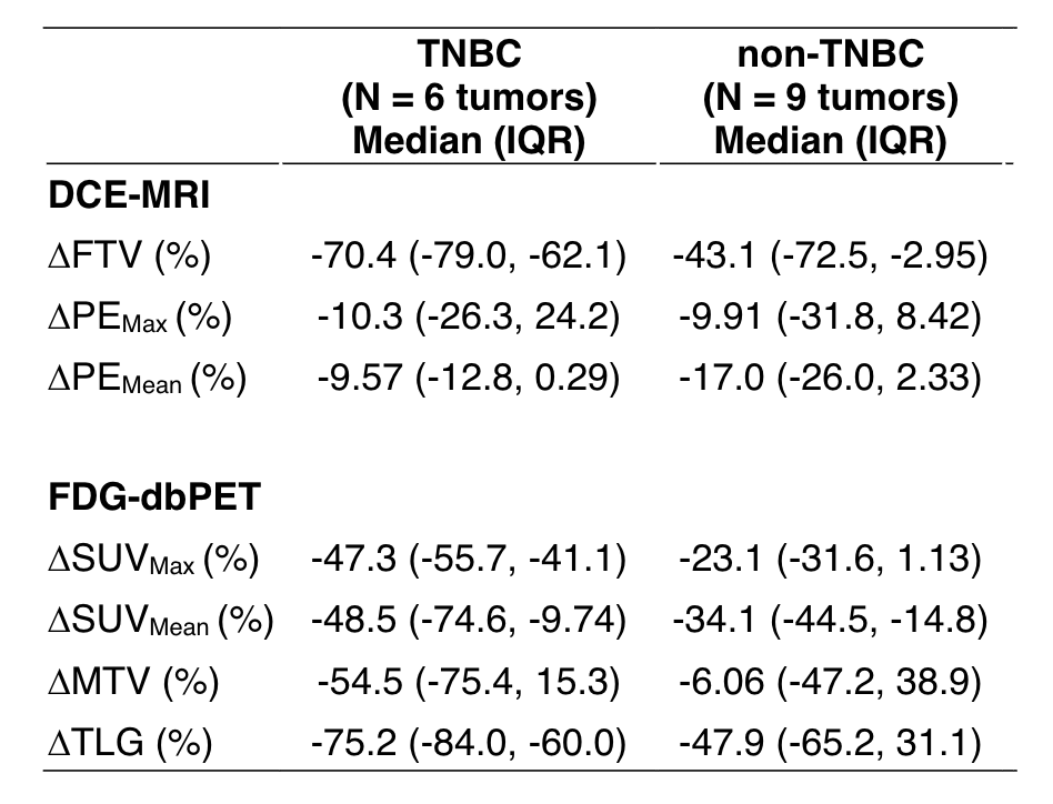
Purpose: Neoadjuvant endocrine therapy (NET) is increasingly used for patients with hormone receptor-positive (HR+) breast cancer. Dynamic contract-enhanced breast MRI is the most accurate modality to monitor tumor response during neoadjuvant chemotherapy (NAC)1, but there is limited research on response to NET.The Endocrine Optimization Protocol (EOP) is a sub-study of the ongoing I-SPY 2 TRIAL testing amcenestrant (an oral selective estrogen receptor degrader [SERD]), with or without addition of abemaciclib (a CDK4/6 inhibitor) or letrozole (an aromatase inhibitor) in patients with stage 2/3, MammaPrint (MP) low-risk (index 0 to 1) or high-risk 1 (index -0.57 to 0), HR+/HER2-negative breast cancer. All I-SPY2 (including EOP) patients undergo MRI at baseline (T0), 3 weeks (T1), 12 weeks (T2), and 6 months, prior to surgery (T3). Functional tumor volume (FTV)2,3 is derived as a quantitative measure of tumor burden from each MRI. A subset of EOP patients also have 3 dedicated breast PET (dbPET) exams with 18F-fluoroestradiol (an estrogen receptor-targeted tracer, FES) at T0, T1, and T3. FES uptake on dbPET indicates the presence of functional estrogen receptor.This study evaluates changes in FTV and FES uptake in patients receiving NET in the ongoing EOP trial. FTV changes in EOP were compared with those in a cohort of patients who received NAC in I-SPY 2.

Methods: The breast MRI and FES-dbPET images from patients in the EOP trial as of June 2022 were evaluated by a blinded central radiology team at a single institution. FTV was measured using standard procedure in I-SPY 2. Percent FTV change (ΔFTV) at Tn (n = 1, 2, or 3) was calculated by 100x(FTVTn-FTVT0)/FTVT0. FES uptake was quantified as standardized uptake value (SUV). Maximum SUV over the tumor volume (SUVmax) was measured using Osirix MD (Pixmeo SARL) and percent change (ΔSUVmax) was similarly defined. For comparison, FTV was evaluated using curated imaging data of I-SPY 2 patients with stage 2/3, MP high-risk 1, HR+/HER2-negative cancer who completed standard NAC between 2010–2016.

Results: We included 55 EOP patients (NET cohort) and 68 I-SPY 2 patients (NAC cohort). At T0, median FTV was 9.8cc for the NET cohort and 10.1cc for the NAC cohort. Table 1 shows the longitudinal FTV change in the two cohorts. At T1, median FTV change was similar in the NET cohort (-33.8%) and NAC cohort (-33.9%). The NET cohort showed a dynamic range of FTV change from -65.4% (1st quartile) to -11.0% (3rd quartile), which covered the 1st to 3rd quartile ranges for the NAC cohort. At T2 and T3, FTV change was more gradual in the NET cohort compared to the NAC cohort.Seven patients in the NET cohort underwent FES-dbPET. At T0, tumor FES uptake exceeded background uptake in all 7 patients with a median SUVmax of 8.2. At T1 and T3, tumor uptake decreased in all patients. Tumor uptake was indistinguishable from background for 3 patients (43%) at T1 and 5 patients (71%) at T3, despite evidence of residual tumor on MRI. The median change of SUVmax was -45.9% at T1 and -74.7% for T3 (Table 2).

Discussion: After 3 weeks of NET, we observed a large dynamic range of FTV change similar to that seen in NAC and a robust decrease in FES uptake. These results suggest the potential for combined use of early MRI change and FES-dbPET to provide scalable biomarkers to stratify response-based NET strategies.

References:

1. Radiology 285: 358–375, 2017

2. Radiology 263:663–672, 2012

3. Radiology 279:44–55, 2016

Abstract No. P5-07-03
2022 San Antonio Breast Cancer Symposium, 8-11 Dec, 2022

The Association Between Symptom Severity and Physical Function among Participants in I-SPY2

Basu A, Umashankar S, Blevins K, Northrop A, Christofferson A, Olunuga E, Cha J, Mittal A, Molina-Vega, Sit L, Brown T, Parker B, Heditsian D, Brain S, Simmons C, Taboada A, Hielen TJ, Ruddy K, Salvador C, Mainor C, Afghahi A, Tevis S, Blaes A, Kang IM, Perlmutter J, Rugo H, Kanaparthi S, Peterson G, Weiss LT, Asare A, Esserman LJ, Melisko M, Hershman D

Background. Patient-reported outcomes (PROs) are increasingly recognized as a valuable component to understand treatment tolerability and toxicity among patients on clinical trials. We have implemented a system for monitoring patient reported outcomes (PROs), symptoms, and quality of life (QOL) using electronic PRO (ePRO) instruments for patients enrolled in the I-SPY2 trial. I-SPY2 is a phase II multi-site clinical trial evaluating the effect of novel neoadjuvant therapies for locally advanced breast cancer. We correlated patient demographic factors with symptoms, investigated the trajectory of symptoms throughout treatment, and sought to characterize symptoms associated with decline in physical function (PF).

Methods. Our study population included 259 I-SPY2 patients that completed surveys on one of 9 study arms (including novel oral taxane/immunotherapy combinations, IV paclitaxel, checkpoint inhibitor+/- LAG3 inhibitor, and control IV paclitaxel +/- trastuzumab/pertuzumab). After the 12 week period of investigational agents, most patients received standard adriamycin and cyclophosphamide (AC). Symptom severity, frequency, and interference was assessed weekly using 33 items from the PRO-CTCAE item bank. PF was assessed using the NIH PROMIS instrument and was evaluated at baseline, inter-regimen (after 12 weeks of treatment), pre-surgery, and 1 and 6 months at follow-up. An odds ratio was used to assess univariate associations between age and race, and symptoms. Regularized multi-variate regression was used to evaluate early symptoms (prior to week 6) predictive of a clinically significant (>5 point T-score) decline in PF from baseline to post-treatment follow-up among all races and age groups.

Results. Of 259 patients (mean age (SD) = 46.8 (13.6)), 160 (58%) were White, 13 (5%) were Asian, 26 (10%) were African American (AA), 25 (9.3%) were Hispanic, and 35 (13.5%) self-reported “Other”. At baseline, AA patients had a higher severity of joint pain than White patients (OR = 14.9, P < 0.05). During study treatment with paclitaxel and/or novel agent within the first 12 weeks of treatment, AA patients and non-white (NW) patients were more likely to report severe vomiting than White patients (OR =13.22 and 12.72, P< 0.05 and P< 0.03 respectively). During treatment with AC, NW patients were more likely to report higher severity of neuropathy than White patients (OR = 5.43, P< 0.03). Among all patients, in analysis of early symptoms predictive of a clinically significant decline in PF between baseline and 1 month post treatment, predictors included high frequency of diarrhea, severity of itching, and severity of joint pain. Further analysis of symptom trajectories revealed that frequency of diarrhea reported rose sharply between baseline and Cycle 2 with 9 patients (7%) reporting occasional or frequent diarrhea to 39 patients (28%) reporting occasional to almost constant diarrhea and remained stable at that proportion for the remainder of treatment. Frequency of diarrhea declined slightly during AC (17%) and dropped to baseline levels by follow-up. In contrast, severity of joint pain persisted post-treatment, rising consistently from baseline through follow- up with 3 patients (2%) reporting moderate to severe joint pain at baseline to 18 patients (35%) reporting moderate to severe joint pain at follow-up.

Conclusion. Among I-SPY2 participants, when higher grade of diarrhea is persistent (or uncontrolled), it impacts physical function even after end of therapy. In some cases, race was also a determinant in symptom trajectory, although a higher enrollment of AA and NW patients will enable more robust estimates to be computed. While some of these early symptom predictors are transient and resolve by the time of follow-up, others persist long-term and contribute more directly towards impaired physical function at follow-up.

Abstract No. P5-05-05
2022 San Antonio Breast Cancer Symposium, 8-11 Dec, 2022

Monitoring for response and recurrence in neoadjuvant-treated hormone receptor-positive HER2-negative breast cancer by personalized circulating tumor DNA testing

Magbanua MM, Rugo H, Brown-Swigart LA, Ahmed Z, Hirst GL, Wolf DM, Lu R, Kalashnikova E, Renner D, Rodriguez A, Liu MC, Yau C, Esserman LJ, van ‘t Veer L, DeMichele A

Background: The detection of circulating tumor DNA (ctDNA) may serve as an early predictor of response and recurrence. In this study, we used a tumor-informed ctDNA test to monitor clinical outcomes in patients with high-risk hormone receptor-positive HER2-negative (HR+HER2-) tumors who received neoadjuvant chemotherapy (NAC) on the I-SPY 2 trial (NCT01042379).

Methods: We collected blood samples at pretreatment, during (at 3 and 12 weeks after initiation of paclitaxel-based treatment with or without an investigational drug), after NAC prior to surgery, 4 weeks after surgery, and annually until clinical diagnosis of recurrence. Cell-free DNA was isolated from plasma (N=329 samples) and ctDNA was detected using a personalized, tumor-informed multiplex polymerase chain reaction next generation sequencing-based test (SignateraTM). All patients were at high risk for recurrence by MammaPrint. The response endpoints were pathologic complete response (pCR) and residual cancer burden (RCB), and the survival endpoint was event-free survival (EFS).

Results: This analysis included 66 patients with HR+HER2- breast cancer who had blood samples collected before, during, after NAC and had at least one blood sample after surgery with sufficient plasma for analysis. 57.1% (32/56) had grade III disease; 72.4% (42/58) were node-positive; 36.2% (21/58) had T3/T4 disease; and 33.3% (22/66) were MammaPrint High 2. The percent ctDNA positivity rates at pretreatment, after NAC prior to surgery, and 4 weeks after surgery were 79.7% (47/59), 6.5% (4/62), and 2% (1/50), respectively. Significantly higher ctDNA positivity rates at pretreatment were observed in patients with larger tumors (95% in T3/T4 vs. 69% in T1/T2, Fisher’s exact p=0.0387), higher grade tumors (94% in Grade III vs. 67% in Grade I/II, p=0.0147) and by MammaPrint score (100% in High 2 vs. 71% in High 1, p=0.0052). In this high-risk HR+/HER2- cohort, 10/66 (15.2%) achieved pCR/RCB 0, who were all ctDNA-negative at surgery. 56/66 (84.8%) had no-PCR, with RCB I (limited residual cancer), II (moderate) and III (extensive) in 7 (10.6%), 31 (47.0%) and 18 (27.3%), respectively. ctDNA-positivity after paclitaxel-based treatment was significantly associated with RCB II/III status (Fisher’s exact p=0.01). All patients in this cohort with persistent ctDNA subsequently had RCB II or III at surgery. 47 patients had paired samples collected after NAC prior to surgery and at 4 weeks after surgery. Of the 47, 91.5% (43/47) were ctDNA-negative at both time points and 8.5% (4/47) were discordant; 1 was ctDNA-negative and later tested ctDNA-positive, while 3 were ctDNA-positive and later tested ctDNA-negative. 61/66 patients had EFS data with a median of 1.6 years of follow up (range: 0.6 to 5.6). 5 tested ctDNA-positive in at least one time point after surgery. Of these, 2 experienced a recurrence (one local relapse and one distant metastasis) and both tested positive at the time of recurrence. For the patient who developed a distant recurrence it was the only blood sample available at a follow-up time point; for the patient who developed a local recurrence, blood from two earlier follow-up time points had tested negative. To date, no recurrences have been observed in those whose test(s) after surgery were negative for ctDNA.

Conclusions: The persistence of ctDNA during neoadjuvant therapy is associated with the extent of residual disease in a cohort of patients with HR+HER2- breast cancer in the I-SPY 2 trial and thus may be useful in identifying patients who are not having an optimal response to therapy. I-SPY 2.2 will test whether ctDNA has utility in redirecting therapy to improve surgical outcome and subsequent prognosis.

Abstract No. P4-02-10
2022 San Antonio Breast Cancer Symposium, 8-11 Dec, 2022

MRI models by response predictive subtype for predicting pathologic complete response

Li W, Onishi N, Wolf DM, Newitt DC, Yau C, Wilmes LJ, Gibbs JE, Price ER, Joe BN, Kornak J, LeStage B, I–SPY2 Imaging Working Group, I-SPY2 Consortium, Esserman LJ, van ‘t Veer L, Hylton NM

Background: MRI predictive modeling is used in the I-SPY 2 neoadjuvant clinical trial as a key component of the pre-RCB (Predicted Residual Cancer Burden) clinical workflow for re-directing “good responders” to skip AC (anthracycline) and proceed to surgery early. The current MRI model is hormone receptor (HR)- and human epidermal growth factor receptor 2 (HER2)-specific, and was trained retrospectively using data from 990 patients in I-SPY 2. Recently, new breast cancer subtypes based on gene expression and pathologic response were proposed by Wolf et al [1]. Their study predicted that drug allocation by the new response-predictive subtype (RPS) would lead to a higher pathologic complete response (pCR) rate than allocation based on HR/HER2 subtypes. In this project, we evaluated the MRI model optimized by RPS and compared it with the HR/HER2 optimized model.

Methods: A total of 990 patients enrolled in I-SPY 2 and randomized to one of 9 drug arms or control were evaluated in this analysis. Functional tumor volume (FTV) was calculated from dynamic-contrast enhanced MRI [2] performed pretreatment (T0), after 3 weeks of treatment (T1), and between sequential drug regimens (T2). pCR was assessed at surgery after treatment was completed. HR/HER2 subtype was defined by HR and HER2 +/-, which resulted in four subtypes: HR+/HER2-, HR+/HER2+, HR-/HER2+, and HR-/HER2- (triple negative). RPS subtype was defined by immune, DNA repair deficiency (DRD), HER2, and BluePrint (BP) subtype (Agendia) biomarkers to define five subtypes: HER2-/Immune-/DRD-, HER2-/Immune+, HER2-/Immune-/DRD+, HER2+/BP-HER2_or_Basal, and HER2+/BP-Luminal. A logistic regression model using at least 1 FTV variable (value at T0, percent change at T1 or T2) was analyzed for predicting pCR. AUC (area under the receiver operating characteristic curve) was used to identify the optimal logistic regression model (highest AUC) in each biomarker-defined subset. For multi-predictor analysis, 10-fold cross validation was used.

Results: 854 patients (301 pCRs, 35%) with FTV evaluations at T0, T1, and T2, HR/HER2 and RPS subtypes, and pCR outcomes were included. Numbers of patients and pCR rates in individual subtypes are listed in Table 1. Of FTV variables, percent change at T2 was selected for inclusion in almost all subtype specific optimal models except HR+/HER2+. FTV at T0 (pretreatment tumor volume) was included in triple negative, HER2-/Immune+, and HER2+/ BP-HER2_or_Basal models. Using the current HR/HER2-specific model, the highest AUC (0.74) was found in triple negatives and the lowest AUC (0.68) was in HR+/HER2+. Using the proposed RPS-specific model, the highest AUC (0.84) was found in HER2-/Immune-/DRD+ and the lowest AUC (0.59) was found in HER2+/BP-Luminal cohorts. Table 1 shows AUCs estimated using predictions generated by HR/HER2- versus RPS-specific models, in the full cohort and in individual HR/HER2 sub-cohorts. AUCs were improved when RPS-specific models were used in full and in HR+/HER2-, HR+/HER2+, and triple negative cohorts. No improvement was observed in the HR-/HER2+ cohort where 97% (72/74) were HER2+/BP-HER2_or_Basal.

Conclusion: Improved prediction of pCR was observed using the RPS-specific MRI model compared to the current HR/HER2-specific model. A new preRCB workflow is being developed to combine MRI-based prediction with core biopsy assessment to re-direct “good responders” to surgery earlier and more precisely based on a patient’s biological subtype.

Abstract No. PD11-01
2022 San Antonio Breast Cancer Symposium, 8-11 Dec, 2022

Evaluation of the PD-1 Inhibitor Cemiplimab in early-stage, high-risk HER2-negative breast cancer: Results from the neoadjuvant I-SPY 2 TRIAL

Stringer-Reasor E, Shatsky RA, Chien J, Wallace A, Boughey JC, Albain KS, Han HS, Nanda R, Isaacs C, Kalinsky K, Mitri Z, Clark AS, Vaklavas C, Thomas A, Trivedi MS, Lu J, Asare S, Lu R, PItsouni M, Wilson A, Perlmutter J, Rugo H, Schwab R, Symmans WF, Hylton NM, van ‘t Veer L, Yee D, DeMichele A, Berry D, Esserman LJ, I-SPY Investigators

Background: I-SPY2 is a multicenter, phase 2 trial using response-adaptive randomization within biomarker subtypes defined by hormone-receptor (HR), HER2, and MammaPrint (MP) status to evaluate novel agents as neoadjuvant therapy for high-risk breast cancer. The primary endpoint is pathologic complete response (pCR). Cemiplimab (Cemi) is a PD-1 inhibitor approved for the treatment of NSCLC, cutaneous basal, and squamous cell cancer. Here, we report current efficacy rates of Cemi in combination with paclitaxel followed by AC.

Methods: Women with tumors ≥ 2.5cm were eligible for screening. Only HER2 negative (HER2-) patients were eligible for this treatment; HR positive (HR+) patients had to be MP high risk. Treatment included paclitaxel 80 mg/m2 IV weekly x 12 and Cemi 350 mg IV given q3weeks x 4, followed by doxorubicin/cyclophosphamide (AC) every 2 weeks x 4. The control arm was weekly paclitaxel x 12 followed by AC every 2-3 weeks x 4. All patients undergo serial MRI imaging; and imaging response (at 3 weeks, 12 weeks and prior to surgery) were used along with accumulating pCR data to continuously update and estimate pCR rates for trial arms. Analysis was modified intent to treat. Patients who switched to non-protocol therapy count as non-pCR. The goal is to identify (graduate) regimens with ≥85% Bayesian predictive probability of success (i.e. demonstrating superiority to control) in a future 300-patient phase 3 neoadjuvant trial with a pCR endpoint within responsive signatures. Cemi was eligible to graduate in 3 pre-defined signatures: HER2-, HR-HER2-, and HR+HER2-. To adapt to changing standard of care, we constructed “dynamic controls” comprising ‘best’ alternative therapies using I-SPY 2 and external data and estimated the probability of Cemi being superior to the dynamic control.  

Results: 60 HER2- patients (28 HR+ and 32 HR-) received Cemi arm treatment. The control group included 357 patients with HER2- tumors (201 HR+ and 156 HR-) enrolled since March 2010. Cemi graduated in HR-/HER2- signature. Estimated pCR rates (as of June 2022) are summarized in the table.Immune-related endocrine disorders include: hypothyroid (14.5%), adrenal insufficiency (10%), hyperthyroid (4.8%),) and thyroiditis (3.2%). Only one grade 3 adrenal insufficiency was observed. All immune related AE’s were manageable. Additional biomarker analyses are ongoing and will be presented at the meeting. Response predictive subtypes (Immune+ vs Immune-) and additional predictive biomarkers were assessed. Associations with pCR will be presented at SABCS.

Conclusion: The I-SPY 2 study aims to assess the probability that investigational regimens will be successful in a phase 3 neoadjuvant trial. Anti-PD-1 therapy with Cemi resulted in a higher predicted pCR rate in HR-/HER2- 55 rate% disease compared to control at 29%. Immune-mediated AE’s were observed. This data is consistent with previously published data using check point inhibitors in early-stage HR-/HER2- breast cancer.

Abstract No. P3-09-01
2022 San Antonio Breast Cancer Symposium, 8-11 Dec, 2022

Characterization of residual disease after neoadjuvant selective estrogen receptor degrader (SERD) therapy using tumor organoids in the I-SPY Endocrine Optimization Protocol (EOP)

Rosenbluth J, Bui TB, Warhadpande S, Phadatare P, Eini S, Bruck M, Molina-Vega J, Pullakhandam K, Schindler N, Brown-Swigart LA, Yau C, Hirst G, Mukhtar R, Giridhar KV, Olopade OI, Kalinsky K, Ewing CA, Wong JM, Alvarado MD, van ‘t Veer L, Esserman LJ, Chien J

Background: Treatment of estrogen receptor (ER)-positive breast cancer with selective estrogen receptor degraders (SERDs) frequently results in the loss or reduction of ER expression. Whether these changes are due to on-target effects of SERDs degrading ER or arise as a mechanism of tumor resistance with associated changes in cellular phenotypes remains unknown. It is critical to distinguish between these possibilities to accurately assess treatment response and determine the most appropriate subsequent therapy. To this end, we created and conducted molecular analyses on patient-derived organoid cultures from post-treatment tissue in patients receiving neoadjuvant SERD therapy for early-stage ER+ breast cancer in the I-SPY2 Endocrine Optimization Protocol (EOP).

Methods: The I-SPY2 EOP study is a prospective, randomized substudy within the I-SPY TRIAL testing the oral SERD amcenestrant alone or in combination with letrozole or abemaciclib in stage 2/3 ER+ Her2-negative breast cancer. Enrollment is ongoing, with patients receiving amcenestrant neoadjuvantly for 6 months until the day before surgery. Tumor tissue is collected at baseline, 3 weeks, and at surgery. Organoids were generated from post-treatment surgical samples. Organoid cultures were optimized based on established methods (Dekkers et al., Nature Protocols, 2021) to assess ER levels and activity. Pre- and post-treatment tissue samples were also assessed for ER, PR, Ki67, and GATA3, a luminal marker and transcription factor that is functionally linked with ER, via immunohistochemistry.

Results: In 7 patients with both pre- and post-treatment tissue samples including fresh surgical samples for organoid generation, the ER in baseline tumor tissue was >=90% in all patients, PR ranged from 40-90%, and Ki67 ranged from 5-30%. In post-treatment surgical tissue from these cases, ER ranged from 0-30%, PR from 0-50%, Ki67 from < 1%-10%, and GATA3 was positive in 5 of 5 cases tested to-date. The creation of organoids from residual disease at surgery was attempted for these 7 patients, with organoids successfully propagated in 5 cases thus far. 3 of 5 organoid cultures were ready for analysis and in all cases strong ER and PR expression in organoids was observed after culture for > 1 month in the absence of amcenestrant. Detailed gene expression profiling (including Mammaprint/Blueprint) and gene set enrichment analyses (GSEA) to assess for intrinsic breast cancer subtype and ER activity in each sample and corresponding organoid culture are in progress and will be reported with the full dataset.

Conclusion: Patient-derived organoid culturing of residual disease after neoadjuvant endocrine therapy is feasible. Neoadjuvant treatment with a SERD can render ER and PR low or absent at the time of surgical resection, which does not necessarily imply the presence of endocrine therapy resistant disease. The use of organoids and additional IHC markers (GATA3) demonstrate that receptor negativity may be an indicator of the drug hitting its target, suggesting ER signaling is still intact. In general, patient-derived tumor organoid cultures modeling residual disease states can be a useful adjunct to existing methods used to monitor the effects of neoadjuvant endocrine therapy and is being explored in the I-SPY EOP trial.

Abstract No. PD5-04
2022 San Antonio Breast Cancer Symposium, 8-11 Dec, 2022

Characterizing the HER2-/Immune-/DNA repair (DRD-) response predictive breast cancer subtype: the hunt for new protein targets in a high-needs population with low response to all I-SPY2 agents

Wolf DM, Yau C, Wulfkuhle J, Gallagher RI, Brown-Swigart LA, Hirst GL, Coppe JP, Magbanua MJM, Sayaman R, I-SPY2 Investigators, Sit L, Hylton NM, DeMichele A, Berry DA, Pusztai L, Yee D, Esserman LJ, Petricoin EF, van ‘t Veer L

Background: In previous work we leveraged the I-SPY2 trial to create treatment response predictive subtypes (RPS) incorporating tumor biology beyond clinical HR/HER2, to better predict drug responses in an expanded treatment landscape that includes platinum agents, dual HER2-targeting regimens and immunotherapy [1]. We showed that best performing schemas incorporate Immune, DRD and HER2/Luminal phenotypes, and that treatment allocation based on these would increase the overall pCR rate to 63% from 51% using HR/HER2-based treatment selection. The RPS schema has been selected for prospective evaluation in I-SPY2. Using the RPS, one would prioritize platinum-based therapy for HER2-/Immune-/DRD+, immunotherapy for HER2-/Immune+, and dual-anti-HER2 for HER2+ that are not luminal. HER2+/Luminal patients have low response rates to dual-anti-HER2 therapy but may respond better to anti-AKT. However, there is still a considerable ‘biomarker-negative’ group of resistant cancers (HER2-/Immune-/DRD-) with very low pCR rates to all tested agents, that require a new therapeutic approach. Here we characterize the protein signaling architecture of these tumors to identify new target candidates.

Methods: 987 I-SPY 2 patients from 10 arms of the trial were considered for this analysis. All have gene expression, pCR and RPS; 944 have distant recurrence free survival (DRFS) data; and 736 have reverse phase protein array (RPPA) data from laser capture microdissected tumor epithelium. These data – known collectively as the I-SPY2-990 mRNA/RPPA Data Resource – were recently made public on NCBI’s Gene Expression Omnibus [GEO: GSE196096]. We focus on HER2-/Immune-/DRD- tumors, applying Wilcoxon and t-tests to identify phosphoproteins that differ between HR+HER2-/Immune-/DRD- and other HR+HER2- tumors; and between TN/Immune-/DRD- and other TNs. The Benjamini-Hochberg (BH) method is used to adjust p-values for multiple hypothesis testing. In addition, the Kaplan-Meier method is used to estimate DRFS.

Results: 201/736 I-SPY 2 patients with RPPA data are classified HER2-/Immune-/DRD- (HR+HER2-: n=138; TN: n=63). Of these, 8.5% (17/201) achieved pCR. Non-responding HER2-/Immune-DRD- had worse outcomes than responders (~75% vs. ~95% DRFS at 5 years). 60/139 phospho-proteins differ significantly between HR+HER2-/Immune-/DRD- and other HR+HER2- tumors (n=122). These tumors are relatively ‘cold’, in that 90% (54/60) of the phosphoprotein activities characterizing this group are at lower levels than in the overall HR+HER2- population; including immune (e.g. pPDL1, pJAK/STAT) and proliferation (e.g., Ki67, CyclinB1, pAURK) endpoints. Phosphoproteins showing higher levels in this subset include ERBB2 (BH p=1.7E-06), Cyclin D1 (BH p=1.4E-05), pAR (BH p=1.4E-05), and ER (BH p=3E-04). Within the TN subset, only 3/139 phospho-proteins differed significantly between TN/Immune-/DRD- and other TN tumors (n=189). These were all immune-related (pPDL1, pSTAT1, and HLA DR), with lower expression in the TN/Immune-/DRD- group.

Conclusion: HR+HER2- and TN patients who are Immune-Low and DRD-Low have very low pCR rates to all tested therapeutics in I-SPY2 including standard chemotherapy, platinum, and immunotherapy. Senolytics (possibly targeting Cyclin D1), HER2low agents, and AR modulators may overcome resistance in HR+HER2-/Immune-/DRD-, whereas an immune activator beyond checkpoint inhibition is suggested for TN/Immune-/DRD- patients.  

[1] Wolf et. al., Redefining Breast Cancer Subtypes to Guide Treatment Prioritization and Maximize Response: Predictive Biomarkers across 10 Cancer Therapies. Cancer Cell 2022

Abstract No. PD5-02
2022 San Antonio Breast Cancer Symposium, 8-11 Dec, 2022

An Organoid Model System to Study Resistance Mechanisms, Predictive Biomarkers, and New Strategies to Overcome Therapeutic Resistance in Early-Stage Triple-Negative Breast Cancer

Bui TBV, Wolf DM, Moore K, Harris IS, Phadatare P, Yau C, Brown-Swigart LA, Esserman LJ, Coppe JP, Wulfkuhle J, Petricoin EF, Campbell M, Selfors LM, Dillon DA, Overmoyer B, Lynce F, van ‘t Veer L, Rosenbluth J

Background: While new treatments and improved subtyping schemas are anticipated to improve treatment response in triple-negative breast cancer (TNBC) patients, therapeutic resistance remains a significant challenge. Moreover, there is an urgent need for additional research model systems to study resistance and residual disease in breast cancer, including aggressive subtypes of breast cancer. Organoid culture is a promising technology used for growing breast cancer cells with high efficiency; however, the extent to which treatment resistance can be modeled using this system is unknown. This research used patient-derived organoid cultures in the context of computational analyses of large molecular and clinical datasets to study resistance mechanisms, biomarkers, and alternative treatment strategies to overcome drug resistance in early-stage TNBC.

Methods: Organoid cultures were derived from breast tumor samples (taken from lumpectomy, mastectomy, or core biopsy samples), digested to the organoid level using collagenase, and grown in three dimensional cultures using a basement membrane extract and a fully-defined organoid medium (Dekkers et al. Nat Protoc 2021). An evaluation of all available I-SPY2 biomarker data (Wolf et al. Cancer Cell 2022) was performed focusing on genes, proteins, and pathways associated with resistance. These were then used to study whether resistance biomarkers identified in patient tumors are also present in organoids propagated from breast cancer post-treatment residual disease. To this end, bulk RNA sequencing data of organoids were normalized and merged with the TCGA dataset (Hoadley et al. Cell 2018) to enable analysis in a larger context, and immunofluorescence staining of organoids was performed. A high-throughput 386 anti-cancer drug compound screen and subsequent synergy testing with the most promising compounds were performed to identify and predict alternative treatment strategies. Additional assays to explore kinome activity in this organoid model are in progress.

Results: A TNBC organoid biobank was established (n=31), which was enriched for inflammatory breast cancer (IBC; n=15), an aggressive form of breast cancer. Most organoids were derived from residual disease after neoadjuvant therapy. Bulk RNA sequencing analysis performed on 10 TNBC organoids revealed 3 subsets that were characterized predominantly by either normal-like/luminal androgen receptor or basal-like features or expressed distinct gene expression profiles, with IBC cases present in all 3 subsets. Intriguingly, the IBC organoids were characterized by higher expression of a number of immune-related signatures, despite an absence of clear immune cells in culture. A residual disease IBC/TNBC organoid resistant to chemotherapy was used to perform the 386-drug compound screen. The organoid model showed resistance to veliparib-cisplatin, consistent with the expression of gene/protein biomarkers predictive of drug resistance found in I-SPY2 (low PARPi7 levels and high pFOXO1 and pMEK1/2 expression). In addition, the screen identified multiple classes of inhibitors as promising synergistic/additive candidates for overcoming resistance to cisplatin.

Conclusion: In this proof-of-principle study, we demonstrated the utility of matching I-SPY2 resistance biomarkers and signatures to residual disease tumor organoid cultures. We show that tumor organoid cultures modeling drug resistance states are a useful complement to existing research models of breast cancer and can be used for compound testing. We have developed a pipeline to propagate residual tumors from patients enrolled in I-SPY2 into organoid cultures to create a broader platform for preclinical drug testing informed by tumor biology with the ultimate goal of enabling faster, more successful translational studies and increased treatment options for resistant disease.

Abstract No. 591
2022 ASCO Annual Meeting, 3-7 Jun, 2022

Improved pathologic complete response rates for triple-negative breast cancer in the I-SPY2 Trial

Yee D, Shatsky RA, Yau C, Wolf DM, Nanda R, van ‘t Veer L, Berry DA, DeMichele A, Esserman L, I-SPY2 Consortium
 

Background: The I-SPY2 Trial evaluates multiple investigative agents in neoadjuvant breast cancer therapy with the primary endpoint of estimated pathologic complete response (pCR) rate. As a platform phase 2 trial it utilizes an adaptive design to compare new regimens with control chemotherapy (weekly paclitaxel followed by AC).

Methods: Specific regimens are assigned based on clinically relevant signatures, including triple negative breast cancer (TNBC). Drug regimens graduate from the trial when the predicted pCR rate in any signature meets the pre-specified threshold of 85% probability of success in a hypothetical 300-patient, 1:1 randomized, phase 3 trial. The strong correlation between pCR rate and event free survival has been reported. To establish the benefit of administering investigational agents in combination with control weekly paclitaxel x 12 in TNBC, we report estimated pCR rates for the first 7 investigational agents.

Results:TNBC accounted for 37% (363/987) of enrolled patients. Only veliparib and carboplatin (VC) and pembrolizumab (Pembro) met the graduation criteria for TNBC. However, compared to control chemotherapy, each drug tested in TNBC resulted in a numerically superior pCR rate compared to control. These findings imply that stratification of TNBC by response-predictive biomarkers may lead to improved pCR rates. For example, we have used gene expression profiling to further refine TNBC classification into Immune enhanced (Immune+), Immune-/DNA Repair Deficient (DRD)+, and Immune-/DRD- classes. TNBC identified as immune enhanced (63%) have an 89% pCR rate to pembrolizumab, while VC is less effective with pCR rate of 71%. Similarly, Immune-/DRD+ (11%) identifies TNBCs with a 80% pCR rate to VC, while pembrolizumab’s pCR rate in this group is only 33%. For tumors that are neither immune enhanced or DRD-positive (Immune-/DRD-; 25%) show numerically improved pCR rates for neratinib (20%), MK2206 (25%), ganitumab (24%), and ganetespib (22%) compared to control (12%). pCR rates for VC (10%) and pembrolizumab (20%) in this group were similar to drugs that did not graduate. For TNBC, many agents in I-SPY2 showed numerically improved pCR rates compared to conventional chemotherapy even when they did not meet our specified definition of graduation.

Conclusions: Further refinement of TNBC signatures should yield improved therapeutic strategies while also sparing women unnecessary systemic therapy.

Clinical trial information: NCT01042379.

Abstract No. 510
2022 ASCO Annual Meeting, 3-7 Jun, 2022

Molecular subtype to predict pathologic complete response in HER2-positive breast cancer in the I-SPY2 trial

Thomas A, Clark AS, Yau C, Wolf DM, van ‘t Veer L, Douglas EH, Chien AJ, Huppert LA, Rugo HS, Shatsky RA, Isaacs C, Berry DA, Yee D, DeMichele A, Esserman L, I-SPY2 Consortium

Background: HER2-positive breast cancer (bc) is a very heterogenous disease. We hypothesized that molecular subtype may predict disease response to investigational agents in HER2+ bc. Here, we report the pathologic complete response (pCR) rate in the first six agents tested in HER2+ bc in the I-SPY 2 trial for the full HER2+ cohort, by molecular subtype, and by disease receptor status.

Methods: Women with HER2+ tumors which were > 2.5 cm were eligible. The I-SPY2 platform trial tests novel agents given neoadjuvantly with a backbone of taxol (T) and trastuzumab (H) followed by doxorubicin and cyclophosphamide. Agents investigated in HER2+ bc were TH (control), MK2206, AMG386, pertuzumab (P), neratinib (N (given in place of H), and TDM1+P (given in place of TH). An investigational arm graduated if there was >85% chance of success compared to control in a 300-person phase 3 neoadjuvant trial. Further details of the I-SPY2 methods have been previously published. Molecular subtyping based on gene expression was utilized to categorize tumors into 5 response predictive subtypes (RPS) (HER2-/Immune-/DRD (DNA repair deficiency)-, HER2-/Immune+, HER2-/Immune-/DRD+, HER2+/Her2_or_Basal and HER2+/Luminal).

Results: For the full HER2+ cohort (N=245) pCR rate was higher in all investigational arms than control (Table). By tumor receptor status, HER2+/HR- tumors (N=89) had a higher pCR rate than HER2+/HR+ tumors (N=156; 63% vs 37%, p = 0.0001). In HER2+/HR- tumors N, MK2206, P and TDM1/P graduated. In HER2+/HR+ tumors P and TDM1/P graduated. 76% (185/245) of I-SPY 2 HER2+ patients were classified as HER2+/Her2_or_Basal and 24% (60/245) were HER2+Luminal. pCR rate was significantly higher in the HER2+/Her2_or_Basal group than in the HER2+/Luminal group (57% vs 15%, p < 0.0001). All agents, except for MK2206, where numbers were small, showed greater efficacy in the HER2+/Her2_or_Basal group than in the HER2+/Luminal group. HER2+/Luminal appeared to be more sensitive to the AKT inhibitor MK2206 than to targeted HER2 agents, though numbers are small.

Conclusions: pCR rates for patients with HER2+ bc treated with investigational agents, particularly dual HER2-blockade, were promising. Molecular response predictive subtype classification provides insight on how to better target therapy. The HER2+/Luminal group had low pCR rates with dual HER2-blockade but may have higher pCR rate with the addition of an AKT inhibitor and identifies a subgroup of HER2+ tumors in need of novel approaches. AKT inhibition for HER2/Luminal is being tested in I-SPY 2.2.

Clinical trial information: NCT01042379.

Abstract No. 504
2022 ASCO Annual Meeting, 3-7 Jun, 2022

Pathologic complete response (pCR) rates for HR+/HER2- breast cancer by molecular subtype in the I-SPY2 Trial

Huppert LA, Rugo HS, Pusztai L, Mukhtar RA, Chien AJ, Yau C, Wolf DM, Berry DA, van ‘t Veer L, Yee D, DeMichele A, Esserman L, I-SPY2 Consortium

Background: Hormone receptor positive (HR+), HER2- breast cancer (BC) is a heterogenous disease. We hypothesized that molecular subtypes capturing luminal, basal, and immune biology could predict response for patients (pts) with HR+/HER2- disease in the I-SPY2 trial.

Methods: I-SPY2 trial is a phase II, randomized, adaptive study evaluating multiple investigational agents as neoadjuvant BC therapy; the primary endpoint is estimated pCR rate. Investigational agents are given with control weekly paclitaxel x 12, followed by AC x 4. Regimens graduate when the predicted pCR rate in any signature meets the pre-specified threshold of 85% probability of success in a hypothetical 300 pt randomized, phase 3 trial. We analyzed estimated pCR rates for the 1st 7 investigational agents in the HR+/HER- subset, analyzed by clinical/molecular features: BluePrint (BP) Luminal vs. Basal, Mammaprint High1 [MP1] vs. Mammaprint High2 [MP2], MP2 is < -0.57, Responsive Predictive Subtype-5 (RPS-5) (classification based on HR, HER2, immune, DNA-repair, and basal/luminal markers), histology, and stage/nodal status.

Results: 38% (379/987) of pts had HR+/HER2- disease. Only pembrolizumab met the pre-specified graduation criteria for HR+/HER2- BC. pCR rates by treatment arm and molecular subtype are described in the Table. 28% were MP2; 72% were MP1. Overall, pCR rates were higher in pts with MP2 vs MP1 disease (30% vs 11%) including with pembrolizumab (55% vs. 21%). 29% were BP Basal, 71% were BP Luminal; BP Basal was more likely to be MP2 than BP Luminal (77% vs 8%). In all arms except MK2206, HR+/HER2- BP Basal pts were more likely to achieve pCR than BP Luminal pts. For MK2206, BP Luminal pts were more likely to achieve pCR. Immune+ by RPS-5 (39% of HR+/HER2-) predicted pCR to pembrolizumab irrespective of BP Basal or Luminal status (11 pCR/16 pts). Results by histology and stage/nodal status will also be reported.

Conclusions: Our data suggest that MP2 and BP Basal signatures identify a subset of HR+/HER2- BC more likely to respond to neoadjuvant therapy; and that an immune signature can identify pts more likely to respond to pembrolizumab. These findings will aid in guiding prioritization of targeted agents with the goal to optimize pCR for all pts.

Clinical trial information: NCT01042379

Abstract No. 592
2022 ASCO Annual Meeting, 3-7 Jun, 2022

Distribution of breast cancer molecular subtypes within receptor classifications: Lessons from the I-SPY2 Trial and FLEX Registry

Cha J, Warner P, Hiatt R, Gomez SL, van ‘tVeer L, Stover-Fiscalini A, Borowsky A, Symmans WF, Wolf DM, Yau C, Yee D, DeMichele A, Berry DA, Esserman L, Audeh W, Modh S, I-SPY2 Consortium

Background: Expression-based molecular subtypes of breast cancer (BC) predict tumor behavior and therapeutic response. Subtype distributions by age and sociodemographics can inform strategies for BC screening, treatment, and prognosis. The conventional approach, adopted by NCI’s Surveillance, Epidemiology, and End Results (SEER) Program, uses HR and HER2 to label: “triple negative” (HR-HER2-), “HER2-enriched” (HR-HER2+), “luminal A” (HR+HER2-), and “luminal B” (HR+HER2+). However, immunohistochemical (IHC)-based receptor labels may not reflect clinically and epidemiologically relevant molecular subtypes that share the same nomenclature, e.g., luminal B.

Methods: We compared IHC labels by HR/HER2 to molecular subtypes by MammaPrint (MP) and BluePrint (BP) for patients in the phase II neoadjuvant I-SPY2 TRIAL for high-risk, stage II-III BC (NCT01042379, n = 981) and in the multicenter, prospective FLEX Registry for stage I-III BC (NCT03053193, n = 5,679).

Results: IHC labels were discordant with MP/BP in 52% of I-SPY2 and 43% of FLEX cases (Table 1). HR-HER2- had the highest concordance with basal-type (99% in I-SPY2, 88% in FLEX). HR+ labels had the least agreement with MP/BP: HR+HER2- tumors were molecularly luminal B and basal in 71% and 29% of I-SPY2 and 40% and 4% of FLEX cases, respectively. HR+HER2+ tumors were molecularly luminal A and HER2-type in 10% and 60% of I-SPY2 and 15% and 36% of FLEX cases, respectively. Of molecularly luminal B cases, only 14% in I-SPY2 and 7% in FLEX were HR+HER2+.

Conclusions: IHC markers collected by population-based registries (SEER) enable BC surveillance. However, IHC labels cannot be used as surrogates for molecular subtypes by MP/BP, especially for luminal B tumors. Given the unmet need to improve management of luminal B BC, we anticipate the growing importance of molecular subtyping to inform treatment and epidemiological research. We propose that the BC research community work with SEER to update its IHC labels to avoid overlap with molecular subtype nomenclature and incorporate such modern classifications when available.

Abstract No. 514
2022 ASCO Annual Meeting, 3-7 Jun, 2022

The ImPrint immune signature to identify patients with high-risk early breast cancer who may benefit from PD1 checkpoint inhibition in I-SPY2

Mittempergher L, Kuilman MM, Barcaru A, Nota B, Delahaye JMJ, Audeh W, Wolf DM, Yau C, Brown-Swigart L, Hirst G, Symmans WF, Lu R, Liu MC, Nanda R, Esserman L, van ‘t Veer L, Glas Annuska, I-SPY2 Investigators

Background: The remarkable increase of novel Immuno-Oncology drugs in many malignancies has led to the need for biomarkers to identify who would benefit. Various predictive biomarkers have been developed (PD-1/PD-L1 expression, mutations in mismatch repair genes and microsatellite instability, tumor mutational burden and immune infiltration), none have consistently predicted efficacy. The I-SPY2 consortium qualified several expression-based immune biology related signatures that predict response to PD1 checkpoint inhibition. Here we assessed whole transcriptome data of high-risk early-breast cancer (EBC) patients who received Pembrolizumab within the neoadjuvant biomarker-rich I-SPY2 trial (NCT01042379), aiming to migrate the I-SPY2 research findings to a robust clinical grade platform signature to predict sensitivity to PD1 checkpoint inhibition.

Methods: Whole transcriptome microarray data were available from pre-treatment biopsies of 69 HER2- patients enrolled in the Pembrolizumab (4 cycles) arm of the I-SPY2 trial. All patients had a High-Risk 70-gene MammaPrint profile. Pathologic complete response (pCR) was defined as no residual invasive cancer in breast or nodes at the time of surgery. Of the 69 patients, 31 had a pCR (12 HR (hormonal receptor)+HER2-, 19 Triple Negative (TN)), while 38 (28 HR+HER2-, 10 TN) had residual disease (RD). To identify the most predictive genes associated with pCR, gene selection was performed comparing pCR and RD groups by iteratively splitting the dataset in training and test, balancing for HR status. Due to limited sample size, leave one out cross validation was used for performance assessment. Genes with effect size > 0.45 were considered significant.

Results: A signature of 53 genes, named ImPrint, was identified with overall sensitivity and specificity > 90% and > 80% for predicting pCR to pembrolizumab in all patients. Sensitivity and specificity in TN were > 95% and ≥70%, and in HR+HER2- > 80% and > 85%, respectively. The Positive Predictive Value (PPV) is 77% for the HR+HER2- subgroup. Biological annotation of the 53 genes showed that over 90% of the genes have known immune system related functions, of which 63% were previously known to be involved in immune response (including genes coding PD-L1 and PD-1, as well as those identified in I-SPY2).

Conclusions: In the signature development phase, ImPrint predicts pCR to Pembrolizumab in a set of 69 high risk EBC with high sensitivity and specificity. The signature features genes with immune-related functions known to be involved in immune response indicating that it might aid identifying patients with an immune-active phenotype. Importantly, ImPrint appears effective in identifying a subset of HR+HER2- patients who could benefit from immunotherapy. External validation in independent dataset(s) is ongoing and will be presented at the time of the meeting.

Abstract No. GS4-02
2021 San Antonio Breast Cancer Symposium, 7-10 Dec, 2021

Analysis of clinical outcomes and expression-based immune signatures by race in the I-SPY 2 trial

Kyalwazi B, Yau C, Olopade O, Chien AJ, Wallace A, Forero-Torres A, Pusztai L, Ellis E, Albain K, Blaes A, Haley B, Boughey J, Elias A, Clark A, Isaacs C, Nanda R, Han H, Yung R, Tripathy D, Edmiston K, Viscusi R, Northfelt D, Khan Q, Sanil A, Berry S, Asare S, Wilson A, Hirst G, Hylton N, Melisko M, Perlmutter J, Rugo H, Symmans F, van’t Veer L, Berry D, Esserman L

Background Transcriptomic immune-related gene signatures have been associated with achievement of pathologic complete response (pCR) and prognosis in the neoadjuvant setting. I-SPY 2 is a multicenter, phase 2 platform trial using response-adaptive randomization within subtypes defined by receptor status (HR/HER2) and MammaPrint (MP) risk to evaluate novel agents as neoadjuvant therapy for women with high-risk breast cancer. Given racial disparities in mortality from breast cancer and the paucity of racial demographic data from clinical trials, we aimed to evaluate the association between racial groups and baseline characteristics, including expression-based subtypes and immune signatures, treatment response, and prognosis of patients enrolled in the I-SPY 2 TRIAL.

Methods Our study population included 990 I-SPY 2 patients. 15 patients identified as part of a racial group with <10 patients enrolled in the trial and were excluded from analysis. Pre-treatment expression data was available for 971 patients. Follow-up data was available for 907 patients; median follow-up time of 4.4 yrs. Chi-square test was used to assess associations between racial groups and pre-treatment SBR grade, HR/HER2 defined subtypes, intrinsic subtype (defined by BluePrint 80-gene molecular subtyping) and residual cancer burden (RCB) class. Logistic regression was used to evaluate race association with pCR. Cox proportional hazard modeling was used to assess the association between racial groups and event free survival (EFS) in a univariate setting, adjusting for pCR status. Association between racial groups and 28 expression signatures related to immune, proliferation, ER and HER2 pathway was analyzed using ANOVA with post-hoc Tukey test in the overall population and in each receptor subtype.

Results Of 975 patients included in our analysis, 787 (81%) were White, 68 (7%) were Asian, and 120 (12%) were Black or African American. No significant associations between race and pre-treatment SBR grade (p=0.49), HR/HER2 defined subtypes (p=0.09), or expression-based subtypes (p=0.25) were observed. pCR rates do not significantly differ by racial groups (Odds ratio of pCR relative to White: 1.00 for Asian and 0.89 for Black or African American); and no significant differences in RCB class distribution by race was observed (p=0.88). Event free survival was not associated with patient racial group in a univariate Cox model (Hazard ratio relative to White: 1.10, p=0.73 for Asian and 1.37, p=0.13 for Black or African American). Among the 28 expression signatures evaluated, four were differentially expressed among racial groups within the overall population (F-test p<0.05): IFN module, B cell signature, Dendritic cell signature, and Mitotic score. Pairwise comparisons between racial groups with post-hoc Tukey test identified significant differences in IFN module expression between Black or African American vs. White (p=0.019) and Dendritic cell signature expression between Asian vs White (p=0.047). Among patients in the TNBC subtype, three signatures (dendritic cell signature, macrophage signature and ERBB2 module) were differentially expressed between Black or African American and White patients (p=0.002, 0.016 and 0.007).

Conclusion Our analysis demonstrates that among women with high risk breast cancer, race does not affect subtype specific response rates nor event free survival. Distribution of subtypes previously shown to be associated with pCR in the I-SPY2 trial did not significantly differ among racial groups indicating race is less likely than tumor biology to predict response. The decreased expression of immune signatures observed in Black or African American women with TNBC suggests possible differential sensitivity to immunotherapy plus combination chemotherapy. Tumor immune multiplex studies are underway to further investigate.

Abstract No. P4-12-02
2021 San Antonio Breast Cancer Symposium, 7-10 Dec, 2021

Improving patient-reported outcome data capture for clinical research: ePRO in ISPY 2, a phase 2 breast cancer study

Northrop A, Christofferson A, Melisko M, Sit L, Olunuga E, Mittal A, Goldman A, Brown T, Heditsian D, Parker B, Brain S, Simmons C, Taboada A, Ruddy KJ, Hieken T, Piltin M Cook K, Salvador C, Mainor C, Afghani A, Tevis S, Blaes A, Kang I, Melin S, Esserman L, Asare A, Hershman DL, Basu A

Introduction: Advances in technology and internet capability have provided an opportunity for efficient collection of Patient Reported Outcomes (PRO) during medical treatment. Here we describe the development and implementation of a system for monitoring patient reported adverse events (AEs) and quality of life (QoL) using electronic PRO (ePRO) instruments for patients enrolled on the Investigation of Serial studies to Predict Your Therapeutic Response with Imaging And moLecular analysis (I-SPY 2 TRIAL), a phase II adaptive platform clinical trial for locally advanced breast cancer.

Methods: We designed an ePRO system to increase the accuracy of patient-reported QoL and AE data collection with the intent to act on symptoms in real time. Using the OpenClinica electronic data capture system, we developed rules-based logic to build automated ePRO surveys, customized to the I-SPY 2 treatment schedule. Weekly surveys contained a maximum of 126 validated, branching logic questions from the Patient Reported Outcomes Measurement Information System (PROMIS®) Health Measures and the National Cancer Institute’s Patient Reported Outcomes – Common Terminology Criteria for Adverse Events (PRO-CTCAE™) instruments. We piloted ePROs at the University of California, San Francisco (UCSF) to evaluate compatibility with a variety of I-SPY 2 patient scenarios (e.g., dose delays). We then staggered rollout of the ePRO system to 22 I-SPY 2 sites to ensure technological feasibility. In order to improve accuracy of data collection, we utilized real-time tracking and developed a Clinical Research Coordinator (CRC) training manual, which integrated workflow diagrams with technical solutions. CRCs were trained using remote video sessions.

Results: The UCSF ePRO pilot began in September of 2020. Over 9-months, we accrued 43 I-SPY 2 patients (average age of 43.8 years), whose interactions with the ePRO system informed design improvements. Of the patients who received a baseline ePRO survey, the completion rate was 75.9% (average age of 44.2 years). This represents an increase from the 15-20% baseline completion rate for the 360 UCSF I-SPY 2 patients who received paper-based PRO surveys between May 2012 – January 2019. As of June 2021, the ePRO system was operational at all 22 I-SPY 2 sites. The UCSF pilot revealed that engagement with patients at critical timepoints improved survey completion. CRCs facilitated patient participation by sending instructional emails and communicating with patients weekly. We tracked data completeness using a Patient Tracking report, which displayed each patient’s survey completion history. This real-time tool enabled CRCs to identify patients who had not completed ePRO surveys prior to their visit, so they could be provided a tablet computer to complete the survey in the clinic. After introducing tablets into the workflow at UCSF, patient completion of the baseline survey increased from 75.9% to 80%.

Conclusion: The transition from paper to electronic QOL and AE data collection improves the ability of patients to complete PRO surveys, but the process must also be optimized and integrated into clinical workflow and trial conduct. In the future, we will present additional results highlighting the feasibility of multilingual ePRO integration into I-SPY 2. ePRO also provides a new opportunity for data analysis, as well as the potential to reduce high grade toxicity through early intervention. It will allow us to assess QoL and AE data by drug regimen, site, provider, and study treatment. The creation of clinician-facing reports also enables access to patient responses in real-time. By implementing ePRO within I-SPY 2, we not only increase efficiency and accuracy of patient-reported data collection, but also improve quality of care and patient safety.

Abstract No. PD9-04
2021 San Antonio Breast Cancer Symposium, 7-10 Dec, 2021

Tumor-released circulating orphan non-coding RNAs reflect treatment response and survival in breast cancer

Goodarzi H, Navickas A, Wang J, Garcia J, Magbanua MJ, Fish L, Brown-Swigart L, Hirst G, Wolf D, Yau C, Chien J, Simmons C, Delson A, Esserman L, van ‘t Veer L

Background: Liquid biopsies have emerged as effective diagnostic tools in disease monitoring and minimal residual disease detection. Circulating tumor DNA (ctDNA) was recently shown to be a predictor of poor response and recurrence in breast cancer. However, ctDNA shedding from breast tumors can rapidly decrease during treatment, resulting in reduced sensitivity in measuring early changes in tumor response or residual cancer burden (RCB) after neoadjuvant chemotherapy (NAC). We recently reported the discovery of orphan non-coding RNAs (oncRNAs), a class of small RNAs that are not present in healthy cells, but emerge from cancer cells. Similar to ctDNA, tumor-released oncRNAs can be used to detect the presence of an underlying tumor; however, since they are actively released by cancer cells, their abundance in the cell-free compartment is substantially higher than ctDNA. Therefore, we hypothesized that monitoring circulating oncRNAs in blood permits a more sensitive approach to measuring treatment response (i.e., pathologic complete response, or pCR) and estimating RCB.

Patients and Methods: Cell-free RNA (cfRNA) was extracted from ~1 ml sera of 72 breast cancer patients treated in the neoadjuvant I-SPY 2 TRIAL with NAC alone or combined with MK-2206 (AKT inhibitor) treatment. For each patient, treatment-naïve samples (T0) were compared with samples from post-treatment and prior to surgery (T3) time-point. RNA samples were subjected to small RNA sequencing (SMARTer), and the presence and abundance of cell-free oncRNA species were then determined by identifying and counting the reads that map to oncRNA loci across samples. Notably, oncRNAs species were pre-annotated from the Cancer Genome Atlas (TCGA), and our approach does not require bespoke personalized assays. We used a machine-learning model to compare abundance of cfRNA species before and after treatment (i.e., T3-T0) to predict pCR and RCB. For this, we split our cohort into a training and a testing set (48 and 24) and trained a model to simultaneously learn the presence of residual disease (pCR vs. no pCR) and its extent (RCB). We then measured the performance of our model on the held-out test data and the entire dataset. To confirm the robustness of our model, we also employed a leave-one-out strategy, whereby pCR and RCBIndex of each patient was predicted using a model that was trained on the other patients in the cohort. Finally, to assess the ability of our oncRNA-based model to risk-stratify patients who fail to achieve pCR (without having been explicitly trained on relapse data), we used the model’s oncRNA score to predict patients at the highest risk of distant recurrence (n=8 out of 36) and performed a multivariate Cox analysis, controlling for HR/Her2 status (median follow-up time was 4.8 years).

Results: The model’s accuracy for predicting pCR—based on changes in circulating oncRNA species between T3 and T0—was 85% for the training data and 79% for the held-out test data (positive predictive value of 75% and negative predictive value of 83%) with combined accuracy of 83%; precision 86% and recall 83%; Pearson R=0.5 for RCB. A leave-one-out strategy showed similar performance (area under ROC of 0.77 versus 0.81 in train-test split). Finally, among the patients who failed to achieve pCR, we observed a significantly higher risk of distant recurrence in those with the highest scores (DRFS: hazard-ratio = 8.4, ANOVA P<0.05).

Conclusion: In this study, we have shown that the changes in tumor-released oncRNA content of the blood are a significant predictor of clinical outcomes. Our results demonstrate that oncRNA fingerprints are blood-accessible, and allow us to build predictive models of tumor response. We are currently expanding this study to additional cohorts, and we expect to report the results for a longitudinal analysis that includes ~200 patients from I-SPY2.

Abstract No. P3-03-07
2021 San Antonio Breast Cancer Symposium, 7-10 Dec, 2021

Single-institution retrospective analysis of lymph node (LN) change on breast MRI in patients with high risk early-stage breast cancer receiving neoadjuvant chemotherapy with and without immunotherapy on the ISPY-2 TRIAL

Christofferson A, Price E, Mukhtar RA, Basu A, Esserman L, Chien AJ

Background: The I-SPY2 TRIAL has shown that the addition of pembrolizumab to standard neoadjuvant chemotherapy results in improved rates of estimated pathologic complete response (pCR) for hormone-receptor positive (HR+) as well as triple negative breast cancer (TNBC). I-SPY2 has tested a number of novel immunotherapy combinations including the combination of pembrolizumab and SD-101, a synthetic oligonucleotide with cytidine-phospho-guanosine (CpG) motifs that stimulates plasmacytoid dendritic cells (pDC) through engagement of toll-like receptor 9 (TLR9). Biomarkers of response to immunotherapy may be different than chemotherapy, and pathologic response may not fully reflect the benefit from checkpoint inhibitors. Early markers of response to immunotherapy are needed to minimize the risk of immune-related toxicity in patients who are unlikely to benefit. The objective of this study is to assess the change in regional lymph nodes (LNs) by breast MRI in I-SPY2 patients receiving neoadjuvant chemotherapy +/- immunotherapy, and to explore if changes in regional LNs correlate with pCR and residual cancer burden (RCB).

Methods: This is a retrospective study of the prospective multicenter I-SPY2 adaptive neoadjuvant trial investigating all patients enrolled at UCSF in 3 study arms between Dec 2015 and April 2021: 1) Control (weekly paclitaxel 80 mg/m2 x 12 weeks followed by AC x 4); 2) weekly paclitaxel + pembrolizumab 200 mg IV every 3 weeks, followed by AC x 4; 3) weekly paclitaxel + every 3 week pembrolizumab + intra-tumoral SD-101 weekly x 4 then every 3 weeks x 2 followed by AC x 4. Serial pre-operative breast MRIs were performed in all patients at baseline, 3 weeks, 12 weeks, and 20 weeks. A single breast radiologist blinded to treatment arm reviewed all MRIs for all patients included in this study and assessed longest diameter and cortical thickness of the largest ipsilateral axillary LN, number and location of abnormal LNs, and development of new abnormal LNs over time. Rates of pCR and RCB were evaluated in patients who showed evidence of LN growth and/or development of new lymphadenopathy (LAD) compared to those who did not. A two-sample test for equality of proportions to measure the statistical significance at alpha=.5 between control and immunotherapy arms and LN change was used.

Results: A total of 43 patients were included, of whom 16 were in the control group, 11 received pembrolizumab, and 16 received pembrolizumab + SD-101. Median age was 45 years, mean tumor size was 4.8 cm, and 60% of tumors were HR+HER2-negative with the remaining 40% being TNBC. Baseline patient and tumor characteristics were similar between the 3 study arms. LN enlargement and/or development of new LAD over time was significantly more common in patients receiving immunotherapy than patients in the control arm (48.1% versus 6.3%, p=0.006). This was seen in both HR+ and TN subtypes. While there was a numerically higher rate of pCR/RCB-1 pathology in patients with increased and/or new LAD compared to those without (64% vs 51%), this difference was not statistically significant. The association of LN change and immune-related toxicities will be reported.

Conclusion: The addition of immunotherapy to standard neoadjuvant chemotherapy in the I-SPY2 trial was associated with an increase in size of ipsilateral axillary LN and/or development of new LAD on serial breast MRI imaging during the course of neoadjuvant treatment. These changes were not associated with worse pathologic response at surgery and should not be assumed to be due to disease progression. Whether these changes could reflect immunotherapy benefit needs to be investigated in a larger trial with longer follow-up.

Abstract No. P3-03-04
2021 San Antonio Breast Cancer Symposium, 7-10 Dec, 2021

Challenges of achieving high image quality on breast MRI for quantitative measurements in the I-SPY 2 TRIAL

Bareng TJ, Gibbs JE, Onishi N, Newitt DC, LeStage B, I-SPY2 Imaging Working Group, I-SPY2 Consortium, Hylton NM

Purpose. To illustrate image quality issues that may impact quantitative measurements used to assess treatment response in a multi-site clinical trial.

Background. Quantitative longitudinal measurements on breast MRI are used to assess patient response to neoadjuvant treatment in the multi-site I-SPY 2 TRIAL. A standardized MR protocol is distributed to sites prior to site initiation. Previous work presented image quality issues1, which may affect measurements that rely on automated computerized methods. Functional tumor volume (FTV), a primary imaging biomarker monitoring treatment response, was more predictive of pathological complete response for protocol adherent exams compared to non-adherent exams2. Image quality may also impact background parenchymal enhancement (enhancement level of normal fibroglandular tissue), which is being investigated as a secondary imaging biomarker. This presentation will show example images demonstrating the challenges of quantitative MR analysis in a multi-site clinical trial.

Image quality issues include:. Motion. Motion due to patient movement during the MRI may result in skin, fat, and breast tissue being poorly defined and blurry. Mis-registration between pre- and post-contrast can cause errors in measurements. Threshold variation. For image processing, two signal intensity thresholds are applied to pixels within the region of interest (ROI), which may be adjusted when less than 50% of the tumor is segmented. The percent enhancement threshold is lowered when poorly enhancing areas of tumor are not segmented. The background threshold is lowered when a bright pixel within the ROI of the pre-contrast images causes relatively darker areas of the tumor to be poorly segmented. Scan duration variation. Scan duration is the time required to scan each T1-weighted acquisition phase. Scan duration variations occur if scan duration is outside the specified protocol range or differs from the scan duration of the patient’s baseline MRI. Longer scan duration may result in overestimation of FTV. Field of View. Field of view (FOV) is the anatomical area being imaged and is directly related to spatial resolution, which plays a key role in the FTV measurement. Incorrect FTV monitoring can occur if FOV is inconsistent between visits, outside the specified protocol range, or is overlarge for the patient.

Discussion. The image quality required for measurements used to assess treatment response in I-SPY 2 differs from the image quality that is acceptable for diagnostic evaluation, including BIRADS category, longest diameter measurements, and visual assessment of washout characteristics. In I-SPY 2, 21 sites use a variety of MRI platforms. Since quantitative measurements are increasingly used to monitor treatment response and guide clinical decision-making, high quality images are essential. Strategies must be implemented to minimize imaging issues and further refine the quantitative measurements. Ongoing studies are examining the impact of image quality factors on accuracy of treatment response prediction.

References. 1.Gibbs J et al. Abstract PS11-08: Operational standardization and quality assurance yield high acceptance rate for breast MRI in the I-SPY 2 TRIAL.

Abstract No. P3-03-02
2021 San Antonio Breast Cancer Symposium, 7-10 Dec, 2021

Diffusion-weighted MRI for prediction of pathologic complete response in HER2- breast cancer treated with pembrolizumab plus neoadjuvant chemotherapy

Li W, Le NN, Onishi N, Newitt DC, Gibbs JE, WIlmes LJ, Kornak J, Partridge SC, LeStage B, Price ER, Joe BN, I-SPY2 Imaging Working Group, I-SPY2 Consortium, Esserman LJ, Hylton NM

Background: Checkpoint blockade pembrolizumab has demonstrated great potential to improve pathologic outcome for HER2- breast cancer. The apparent diffusion coefficient (ADC) is a non-contrast MRI-derived biomarker that is sensitive to changes in tumor cellularity. Clinical trial ACRIN 6698, a sub-study of I-SPY 2, demonstrated that ADC can predict pathologic complete response (pCR). This study compares the utility of ADC for early prediction of pCR in patients with HER2- breast cancer randomized to pembrolizumab versus standard neoadjuvant chemotherapy (NACT) in I-SPY 2.

Methods: A retrospective analysis of imaging and clinical data was performed on a cohort of 249 women diagnosed with high-risk, stage II/III breast cancer. All patients were randomized to the standard NACT (paclitaxel) or pembrolizumab plus paclitaxel for 12 weeks, followed by doxorubicin plus cyclophosphamide. MRI exams performed at pretreatment (T0) and 3 weeks after the treatment started (T1) were analyzed. Tumor ADC was calculated within manually delineated region-of-interests on diffusion-weighted MRI. The percent change of ADC from T0 to T1 was evaluated in the prediction of pCR after NACT. Statistical analysis included Wilcoxon rank sum test and the area under the ROC curve (AUC). A p-value <0.05 was considered statistically significant.

Results: A subcohort of 103 patients with analyzable diffusion-weighted MRI exams and known pCR (n=30)/non-pCR (n=73) outcome were included in this analysis. Among 103 patients, 62 had HR+/HER2- and 41 had triple negative breast cancer. Twenty-eight patients (17 HR+/HER2- and 11 triple negative) were randomized to receive pembrolizumab and 75 (45 HR+/HER2- and 30 triple negative) to standard NACT. Tumor ADC increased at 3 weeks in both standard and pembrolizumab cohorts with median ADC change of 11.5% (interquartile range [IQR]: 4.6, 16.2)% and 14.4% (IQR: 0.2, 19.9)%, respectively. In the pembrolizumab cohort, the difference in ADC change between non-pCR and pCR groups was estimated as -9.7% (95% confidence interval [CI]: -22.4, -0.9), with ADC increasing more in the pCR group. The AUC of predicting pCR in the pembrolizumab cohort was estimated as 0.73 (95%CI: 0.52, 0.93), while it was estimated as 0.63 (95% CI: 0.43, 0.83) in the standard NACT cohort. In comparison, the AUCs using functional tumor volume (FTV) to predict pCR were 0.61 (95%CI: 0.39, 0.83) and 0.66 (95% CI: 0.47, 0.85) in the corresponding cohorts (Table 1). The results suggest that ADC had higher association with pCR than FTV in the pembrolizumab cohort and FTV had higher association than ADC in the standard cohort.

Conclusions: Tumor ADC, measured using diffusion-weighted MRI, demonstrates potential as a biomarker for assessing early response to immunotherapy in the neoadjuvant setting for high risk HER2- breast cancer. This study is limited by sample size. Future analysis with larger cohorts is warranted.

Abstract No.
2021 San Antonio Breast Cancer Symposium, 7-10 Dec, 2021

Functional tumor volume at 3 and 6-week MRI as an indicator of patients with inferior outcome after neoadjuvant chemotherapy

Onishi N, Gibbs JE, Li W, Newitt DC, Price ER, LeStage B, Symmans WF, DeMichele AM, Yau C, I-SPY2 Imaging Working Group, I-SPY2 Consortium, Esserman LJ, Hylton NM

Purpose. Functional tumor volume (FTV) is a quantitative measure of tumor burden derived from dynamic contrast-enhanced breast MRI1, 2. In the I-SPY 2 TRIAL, FTV is measured during neoadjuvant chemotherapy (NAC) at pre-treatment (T0), 3 weeks (T1), 12 weeks (T2), and pre-surgery (T3) time points. In I-SPY2 protocol amendment 18, activated in Dec 2017, an optional MRI at 6 weeks (T1a) recommended at clinician’s discretion was added for patients with low response at T1. Patients are treated with standard NAC with or without addition of experimental agents. A treatment escalation option being planned for future I-SPY2 implementation will give patients with suboptimal response the opportunity to pursue more aggressive therapy. T1 and T1a MRI may be helpful to select candidates for this option. We retrospectively investigated the ability of FTV reduction at 3 and 6 weeks to detect non-responders.

Methods. We included 104 patients who underwent T1a MRI between Jan 2018 and Mar 2021. FTV was measured using in-house software developed in IDL (Exelis Visual Information Solutions, Boulder, CO). FTV reduction at 3 and 6 weeks was dichotomized to under and over with a cutoff of 20% reduction from T0 to T1 and 65% reduction (the 3D equivalent of size-based partial response criteria by RECIST) from T0 to T1a, respectively. Treatment outcome was evaluated based on residual cancer burden (RCB) on surgical pathology, an established surrogate for survival outcome3. Patients with RCB 0/I were considered as responders and those with RCB II/III as non-responders. Fisher’s exact test was used to examine the association between FTV reduction and treatment outcome, with P <0.05 considered statistically significant. Ability of FTV reduction to detect non-responders was assessed by positive predictive value (PPV) and sensitivity, where non-responder was defined as “positive”.

Results. Of the 104 patients, 49 patients (31 HR+HER2–;16 HR–HER2–; 2 HR+HER2+) who had both RCB and analyzable FTVs were included. Other patients were excluded because of missing RCB or FTV data (n = 18) or not having completed the assigned therapy (n = 37).FTV reduction at T1 and T1a was associated with outcome (P = 0.022 and <0.001, respectively) (Table 1). FTV reduction at T1a was also associated with outcome in 26 patients with <20% reduction at T1 (P = 0.047). The combined criteria of <20 % reduction at T1 and <65 % reduction at T1a detected non-responders with PPV of 82% and sensitivity of 95%, which outperformed the T1 only or T1a only criterion (Table 2).Ability of combined criteria was experimentally tested using 20% cutoff for T1 and various cutoffs for T1a (Table 2). Criteria of <20 % reduction at T1 and <50 % reduction at T1a detected non-responders with PPV of 89% and sensitivity of 89%(Table 2).

Conclusion. In this early small study, combined criteria using FTV reduction at 3 and 6 weeks of NAC showed high PPV and high sensitivity in early detection of non-responders as candidates for the treatment escalation option.

Reference. 1. Radiology 263:663–672, 2012. 2. Radiology 279:44–55, 2016. 3. J Clin Oncol. 2007 Oct 01; 25(28) 4414-4422

Abstract No. P3-02-01
2021 San Antonio Breast Cancer Symposium, 7-10 Dec, 2021

Initial experience of FES-dedicated breast PET imaging of early-stage ER+ invasive lobular carcinoma

Jones EF, Hathi DK, Konovalova N, Molina-Vega J, Newitt DC, Lawn-Heath C, Ray KM, Joe BN, Heditsian D, Brain S, I-SPY2 Imaging Working Group, I-SPY2 Consortium, Chien AJ, Esserman LJ, Hylton NM, Mukhtar RA

BACKGROUND: Invasive lobular carcinoma (ILC) is the second most common histological subtype of breast cancer, representing 15% of all invasive breast cancers. Most ILC tumors are estrogen receptor-positive (ER+) and may respond to endocrine therapy. However, tumor biologic factors such as ER functionality, cell proliferation, and molecular traits may influence endocrine treatment response and long-term recurrence risk, thus necessitating a comprehensive approach to characterize the primary breast tumor. [18F]fluoroestradiol (FES) is a radiotracer developed for positron emission tomography (PET) imaging of ER status. For this work, we studied the utility of imaging FES uptake in early-stage primary ER+ ILC lesions, using high-resolution dedicated breast PET (dbPET) to assess the relationship between FES uptake and tumor characteristics.

METHODS: With institutional review board approval, patients with biopsy-proven ER+/HER2- ILC were prospectively imaged using dbPET with 5 mCi of FES before treatment. FES uptake (SUVmax, SUVmean, and SUVpeak), tumor uptake volume (TUV), and background parenchymal uptake (BPU) values were calculated. Background values (SUVbkg) were obtained from the normal region of the ipsilateral breast. Lesions with background-corrected SUVmax 2 times higher than SUVbkg were considered FES-avid. Tumor grade, Ki67 cell proliferation index, and ER expression were obtained from core biopsies before treatment. Ki67 was dichotomized to low and high using a 20% cutoff1. Tumor size (longest diameter) was measured by magnetic resonance imaging (MRI). Spearman rank correlation was used to assess the relationship between FES uptake and tumor size. Differences between FES uptake at high and low Ki67 were compared using a Wilcoxon rank-sum test.

RESULTS: 13 treatment-naïve ILC patients aged 32-82 years were included in this analysis (Table 1). Despite all lesions exhibiting strongly positive ER expression >90% by immunohistochemistry (IHC), we observed varying FES avidity with 9 FES avid and 4 FES non-avid ILC lesions. SUVmax, TUV, and TBR had substantial median differences between Ki67 high and low lesions (5.9, 4.3, and 9.6, respectively), but the difference did not achieve statistical significance. FES tumor uptake also correlated with tumor size, with the highest correlation observed for SUVpeak (ρ = 0.71 (95% CI: 0.22, 0.91), p=0.010) (Table 2).

CONCLUSION: We found that not all highly ER expressing ILC by IHC were FES-avid. As FES-dbPET captures information from the entire tumor, it provides a more comprehensive assessment of functional ER status than IHC of a limited tumor sample. FES uptake in ILC also relates to tumor size and Ki67. This is an ongoing study; additional data may help to guide endocrine therapy decisions. Future studies with a larger cohort are planned to assess the relationship between FES uptake and tumor grade and molecular risk profiles. 1. Acs, B. et al. Ki-67 as a controversial predictive and prognostic marker in breast cancer patients treated with neoadjuvant chemotherapy. Diagn Pathol12, 20, doi:10.1186/s13000-017-0608-5 (2017).

Abstract No. P3-02-02
2021 San Antonio Breast Cancer Symposium, 7-10 Dec, 2021

FES-dedicated breast PET uptake in early-stage ER+ breast cancers

Jones EF, Hathi DK, Molina-Vega J, Newitt DC, Lawhn-Heath C, Ray KM, Joe BN, Heditsian D, Brain S, Mukhtar RA, Chien AJ, Rugo HS, I-SPY2 Consortium, I-SPY2 Imaging Working Group, Esserman LJ, Hylton NM

BACKGROUND: Patients with ER+ breast cancer may have a recurrence risk of aggressive disease. While clinical evidence suggests that ER+ tumors are responsive to endocrine therapy, up to one-third of patients with early-stage ER+ disease may not respond to endocrine therapy. Tumor biologic factors such as ER functionality, cell proliferation, and molecular traits may influence endocrine treatment responsiveness and long-term recurrence risk. More comprehensive tools are needed to depict the primary breast tumor. [18F]fluoroestradiol (FES) is a radiotracer developed for positron emission tomography (PET) imaging of ER status. We used FES with a high-resolution dedicated breast PET (dbPET) to quantify ER expression in primary ER+ tumors and assessed the relationship between FES uptake and tumor characteristics.

METHODS: With IRB approval, patients with biopsy-proven ER+/HER2- breast cancer were imaged using dbPET with 5 mCi of FES before treatment. FES uptake (SUVmax, SUVmean, and SUVpeak), background parenchymal uptake (BPU), tumor uptake volume (TUV), and tumor to background ratio (TBR) were calculated. Background values (SUVbkg) were obtained from the normal region of the ipsilateral breast. Lesions with background-corrected SUVmax 2 times higher than SUVbkg were considered FES avid. Tumor size (longest diameter) was measured by MRI. The histologic subtype, ER expression, tumor grade, and Ki67 were obtained from core biopsies before treatment. Ki67 was dichotomized to low and high using a 20% cutoff. Spearman’s rank correlation was used to assess the correlation between FES uptake and tumor size. Differences between FES uptake, histologic subtype, and Ki67 were compared using a Wilcoxon rank-sum test.

RESULTS: 19 treatment-naïve patients were included in this analysis as part of an ongoing study. Patient and tumor characteristics are listed in Table 1. While all patients had ER positivity >90% by immunohistochemistry (IHC), we observed varying FES avidity in ER+ breast cancers, with 14 FES avid and 5 non-FES avid lesions. There was a statistically significant difference between FES avid vs. non-avid lesions measured by all uptake metrics except BPU. FES uptake in invasive ductal carcinoma was similar to invasive lobular carcinoma. FES uptake correlated with tumor size, with the highest correlation ρ = 0.58, 95% CI (0.17, 0.84), p=0.012, detected in TUV. FES uptake was associated with Ki67, with all uptake metrics except BPU showing a statistically significant difference between high and low Ki67 expression..

CONCLUSION: We found that not all lesions that were highly ER+ by IHC were FES avid. FES-dbPET captures information from the entire tumor, providing a more comprehensive assessment of functional ER status than IHC of a limited tumor sample. Moreover, FES uptake correlates with tumor size and cell proliferation. This is an ongoing study; additional data may help to guide endocrine therapy decisions. Future studies with a larger cohort are planned to assess the relationship between FES uptake and tumor grade and molecular risk profiles.

Abstract No. OT1-10-02
2021 San Antonio Breast Cancer Symposium, 7-10 Dec, 2021

I-SPY2 endocrine optimization protocol (EOP): A pilot neoadjuvant endocrine therapy study with amcenestrant as monotherapy or in combination with abemacicilib or letrozole in molecularly selected HR+/HER2- clinical stage 2/3 breast cancer

Chien AJ, Kalinsky KM, Molina-Vega J, Mukhtar R, Giridhar K, Olopade OI, Basu A, Asare SM, Henderson P, Hirst G, Lu R, Jones E, Hylton N, Brown-Swigart L, van ‘t Veer LJ, Yee D, Mayer I, Esserman LJ

Background: There is no clinical equipoise on the best upfront management of patients with early-stage hormone receptor-positive (HR+)/HER2-negative (HER2-) breast cancer (BC) that is high-risk by clinicopathologic criteria, and low-risk based on molecular profiling. These patients are unlikely to respond to chemotherapy. However, these patients still have risk, often risk of late recurrence, despite standard adjuvant endocrine therapy. Novel endocrine-based strategies that are more effective and tolerable than current standard therapies are needed for this population. Next-generation orally-bioavailable selective estrogen receptor degraders (oSERDs) with improved pharmacokinetic (PK) properties are promising potential therapies for HR+ BC. The oSERD amcenestrant has demonstrated a favorable safety profile and encouraging efficacy in a phase I/II dose escalation and expansion trial for heavily pre-treated patients with HR+ metastatic BC and is an attractive agent for assessment in the neoadjuvant BC setting. The neoadjuvant setting offers a unique opportunity to study novel agents and to assess early biological endpoints. However, one of the challenges in studying endocrine-based strategies in the neoadjuvant setting is the lack of a robust surrogate endpoint to reliably predict response and benefit. The I-SPY2 Endocrine Optimization Protocol (EOP) is a pilot sub-study within the main I-SPY2 TRIAL that will test amcenestrant alone or in combination with abemaciclib or letrozole. EOP will test the feasibility of using the I-SPY2 platform to test novel endocrine-based strategies in the neoadjuvant setting in patients with clinical high-risk, molecular low-risk, HR+/HER2- tumors, and will generate a rich database of imaging, molecular, and pathologic correlative endpoints that may potentially inform the improved assessment of response to neoadjuvant endocrine therapy.

Trial Design/Eligibility/Accrual: The I-SPY2 EOP is a prospective, randomized, open-label trial specifically for patients with HR+/HER2-negative MammaPrint (MP) low-risk tumors that are at least 2.5 cm in size. Eligible patients are identified during the screening process for the parent I-SPY2 trial. The planned total accrual for the EOP is 120 patients. Patients are randomized 1:1:1 to one of 3 oral treatment arms: 1) amcenestrant 200 mg daily; 2) amcenestrant 200 mg daily + abemaciclib 150 mg bid; 3) amcenestrant 200 mg daily + letrozole 2.5 mg daily. Patients are treated for 6 months prior to surgery. Premenopausal women must receive concomitant monthly ovarian suppression. Serial breast MRIs, breast biopsies, blood, and patient reported outcomes (PROs) are being collected, and patients will be followed for 10 years for recurrence and survival. Serial dedicated breast PET (dbPET) scans and PKs will be assessed in a subset of patients.

Objectives/Statistics: The primary objective of the EOP is to investigate the feasibility of enrolling and treating molecularly-selected patients with early stage HR+/HER2- MP low-risk BC in a randomized neoadjuvant trial using an oral-SERD backbone. Treatment will be determined to be feasible if ≥75% of enrolled patients complete ≥75% of assigned study therapy. Secondary objectives include the safety, tolerability, PROs, and PKs related to amcenestrant +/- abemacilcib and letrozole, as well as the assessment of imaging, pathologic, and molecular correlative endpoints as potential biomarkers of response to neoadjuvant endocrine therapy.

Status: This study opened in May 2021. Accrual is ongoing.

Abstract No. PD8-07
2021 San Antonio Breast Cancer Symposium, 7-10 Dec, 2021

Evaluation of Tucatinib + (Paclitaxel + Pertuzumab + Trastuzumab) followed by AC in high-risk HER2 positive (HER2+) stage II/III breast cancer: Results from the I-SPY 2 TRIAL

Potter DA, Roesch E, Yau C, Lu R, Wolf D, Samson S, Stafford D, Albain KS, Isaacs C, Trivedi M, Yee D, Boughey, Thomas A, Chien AJ, Hylton N, Li W, DeMichele A, Perlmutter J, Symmans WF, Hershman DL, Melisko M, van ‘t Veer L, Wilson A, ASare SM, Berry DA, Schwab R, Rugo HS, Esserman LJ

Background: I-SPY 2 is a multicenter, phase 2 trial using response-adaptive randomization within molecular subtypes defined by receptor status and MammaPrint (MP) risk to evaluate novel agents as neoadjuvant therapy for women with high-risk breast cancer. Tucatinib is a potent HER2 (ErbB2) tyrosine kinase inhibitor, selective for HER2 vs. epidermal growth factor receptor (EGFR) and is active vs. brain metastases. Safety and efficacy of tucatinib combined with paclitaxel, pertuzumab, and trastuzumab are unknown and were tested in a planned 10 patient (pt) safety run-in of the I-SPY 2 trial. Methods: Women with tumors ≥ 2.5cm were eligible for screening. Only pts with tumors that were HER2+ by FISH were eligible for this treatment. Treatment included tucatinib (max dose 300 mg) BID for 12 weeks with weekly paclitaxel 80 mg/m2 and trastuzumab (2 mg/kg weekly following loading), and pertuzumab (420 mg every 3 weeks following loading), followed by doxorubicin/cyclophosphamide (AC) every 2 weeks x 4. The control arm was weekly paclitaxel and trastuzumab with pertuzumab for 12 weeks followed by AC every 2 weeks x 4. All pts undergo serial MR imaging and response at 3 & 12 weeks is combined with real time pCR data to estimate, and continuously update, the predicted pCR rate for each trial arm. The goal of the trial is to identify/graduate regimens with ≥85.% Bayesian predictive probability of success (i.e. demonstrating superiority to control) in a future 300-patient phase 3 neoadjuvant trial with a pCR endpoint. This run-in arm was conducted to determine safety of combining tucatinib with paclitaxel/trastuzumab/pertuzumab, monitoring special adverse events of interest including LFT elevations and gastrointestinal toxicities.

Methods: The I-SPY 2 methods have been previously published.

Results: 20 pts were evaluable in tucatinib treatment arm. The control arm included 329 historical controls enrolled since April 2010. The initial tucatinib dose was 300 mg BID. After enrollment of the first 8 pts, there were 3 pts with grade 3 LFT elevations, 2 pts with grade 2/3 diarrhea, 1 pt with grade 2 neutropenia, and 1 pt with grade 3 nausea. After this safety review, the tucatinib dose was lowered to 250 mg BID. Among 5 additional pts enrolled, 3 developed grade 2/3 LFT abnormalities. The protocol was then modified to tucatinib 150 mg BID days 1-28 and then 250 mg BID days 29-84; 7 pts were treated. Safety data were reviewed after 20 pts were enrolled; the arm was then suspended due to similar LFT elevations regardless of tucatinib dose reduction or schedule. 7 of 20 pts (35%) had reversible Grade 3 or higher ALT/AST elevation. No pt met criteria for Hy’s Law. In terms of efficacy, 12 of 14 evaluable pts had > 80% reduction of tumor volume by 12 weeks, measured by MRI assessment of functional tumor volume (FTV).

Conclusion: The goal of the run-in arm was to determine the safety of adding tucatinib to the combination of paclitaxel/trastuzumab/pertuzumab. The addition of tucatinib resulted in unacceptable but reversible LFT elevations despite tucatinib dose reduction. Tucatinib containing therapy resulted in >80% decline in tumor volume at 12 weeks in 86% of pts. Tucatinib showed a high level of activity when combined with paclitaxel/trastuzumab/pertuzumab, but the combination is not feasible. Table: Number of pts with grade 2, 3, and 4 LFT elevations by treatment schedule (highest grade per patient per event, ALT or

Abstract No. P2-01-03
2021 San Antonio Breast Cancer Symposium, 7-10 Dec, 2021

Elucidating the biology of circulating tumor DNA (ctDNA) shedding across receptor subtypes in high-risk early-stage breast cancer

Sayaman RW, Wolf DM, Yau C, Brown-Swigart L, Hirst G, Sit L, O’Grady N, Delson AL, I-SPY2 Investigators, Esserman L, van ‘t Veer LJ, Magbanua MJM

Background: Identifying mechanisms that govern the shedding of ctDNA in blood could inform the use of liquid biopsy in individual patients. Previous studies in the I-SPY2 neoadjuvant trial involving high-risk breast cancer showed that the detection of ctDNA before treatment was associated with aggressive clinical characteristics and residual ctDNA after treatment was associated with poor outcomes. Moreover, ctDNA positivity rates significantly varied across breast cancer subtypes suggesting that ctDNA shedding may in part be driven by subtype-specific etiology. We performed genome-wide transcriptomic analysis to identify genes and biological processes associated with increased ctDNA shedding within and across receptor subtypes.

Methods: Our study involved 227 patients in I-SPY2 with tumor gene expression and ctDNA data at pretreatment. All patients were at high risk for recurrence (MammaPrint high). Each subtype: HR+HER2- (n=109), HER2+ (n=19), and triple negative breast cancer (TNBC, n=99) was evaluated independently. We performed differential expression (DE) analysis on the global transcriptome (m=19,134 genes) and curated gene signature (cGS, m=31 signatures developed in I-SPY2) data between ctDNA+ and ctDNA- patients at baseline. Gene-set enrichment analysis (GSEA) was also performed across hallmark (H, m=50), canonical pathway (CP, m=5,501), gene ontology (GO, m=9,996) and immunologic (IM, m=4,872) gene sets. Features were associated with ctDNA shedding if Benjamini-Hochberg adjusted p< 0.05. For subtypes with smaller sample size and unbalanced groups, we also report features with nominally significant p< 0.05.

Results: ctDNA positivity rate was significantly higher in TNBC (91%) than in HR+HER2- and HER2+ (65% and 74% respectively, Fisher p<0.001). The HR+HER2- subtype had the most significant hits for DE analysis between ctDNA+ and ctDNA- patients, with 0.2% of genes and 3.2% of cGS. No genes or cGS were differentially expressed in TNBC and HER2+, likely due to imbalance or small size of these groups. For GSEA, we observed the most significant number of enrichments in HR+HER2- subtype, with 58%, 21.8%, 4.4% and 40.3% of H, CP, GO, and IM gene sets enriched, respectively. In the HER2+ subtype, 40% H, 15.7% CP and 36.4% IM gene sets were significantly enriched, while no gene sets were enriched in TNBC. To identify common mechanistic themes across subtypes, we also considered nominally significant features in DE and GSEA. Processes associated with infection and innate immune responses were enriched in ctDNA+ patients, while adaptive immune response and antigen presentation—e.g., T-cell, TCR and MHC II protein complex, and downregulation of MYC targets were enriched in ctDNA- patients. HR+HER2- and HER2+ subtypes shared the most common modulated features with 134 genes and 2,165 gene sets, including up-regulation of cell cycle and proliferation in ctDNA+ patients, as well as up- or down-regulation of specific immunologic and metabolic processes. In contrast, TNBC gene set enrichment was associated with more distinct biologic processes, sharing common enrichment of 113 and 27 gene sets with HR+HER2- and HER2+ subtype, respectively.

Conclusions: Findings from our exploratory analysis suggest a key role of immune response pathways in the control of ctDNA release. Additionally, tumor cell proliferation was associated with increased shedding in HR+HER2- and HER2+ subtypes, while down regulation of MYC targets was associated with ctDNA- patients across all subtypes. These suggest an important role of cell cycle in ctDNA shedding. Overall, our analysis revealed common and unique mechanisms potentially associated with ctDNA shedding across and within subtypes. However, due to the unbalanced groups and limited sample sizes, validation in a larger cohort is warranted.

Abstract No. 508
2021 ASCO Annyal Meeting, 4-8 Jun, 2021

Evaluation of intra-tumoral (IT) SD-101 and pembrolizumab (Pb) in combination with paclitaxel (P) followed by AC in high-risk HER2-negative (HER2-) stage II/III breast cancer: Results from the I-SPY 2 tria

Chien AJ, Soliman HH, Ewing CA, Boughey JC, Campbell MJ, Rugo HS, Wallace AM, Albain KS, Stringer-Reasor EM, Church AL, Kalinsky K, Elias AD, Mitri ZI, Clark AS, Nanda R, Thomas A, Yau C, I-SPY2 Consortium, Berry DA, Esserman L

Background: I-SPY 2 is a multicenter, phase 2 trial using response-adaptive randomization within molecular subtypes defined by receptor status and MammaPrint (MP) risk to evaluate novel agents as neoadjuvant therapy for women with high-risk breast cancer. SD-101 is an investigational Toll-like receptor 9 (TLR9) agonist CpG-C class oligodeoxynucleotide that stimulates the production of IFN-α and interleukin (IL)-12, functional maturation of plasmacytoid dendritic cells, and production of cytotoxic antibodies. IT SD-101 was combined with systemic anti-PD-1 antibody Pb to investigate the antitumor and immunologic activity of this novel immunotherapeutic strategy.

Methods:Women with tumors ≥ 2.5cm were eligible for screening. Only pts (pts) with HER2- disease were eligible for this treatment. Treatment included weekly P x 12 in combination with IT SD-101 2 mg/ml (1 ml for T2 tumors, 2 ml for >T3 tumors) weekly x 4, then q3 weeks x 2, and IV Pb q3 weeks x 4, followed by doxorubicin/cyclophosphamide (AC) q2-3 weeks x 4 (SD-101+Pembro 4). Pts in the control arm received weekly P x 12 followed by AC q2-3 weeks x 4. The I-SPY 2 methods have been previously published. This investigational arm was eligible for graduation (>85% chance of success in a 300-person phase 3 neoadjuvant trial) in 3 of 10 pre-defined signatures: HER2-, hormone receptor (HR)+/HER2- and HR-/HER2-.

Results: 75 pts were randomized and evaluable in SD-101+Pembro 4 treatment arm. The control arm included 329 historical controls enrolled since April 2010. The study arm was stopped due to maximal patient accrual. Pt characteristics were balanced; 56% HR+, 44% HR-. The probability that SD-101+Pembro4 was superior to control exceeded 97% for all eligible tumor signatures, but did not reach the threshold for graduation in any of the signatures. However, it is notable that the rate of pCR/Residual Cancer Burden 1 (RCB1) in the HR+/HER2- signature was 51%. Preliminary safety events for SD-101+Pembro 4 include increased rates of fever, neutropenia, febrile neutropenia, transaminitis, and immune-related events, including adrenal insufficiency.

Conclusions: The SD-101+Pembro 4 regimen was active but did not meet the pre-specified threshold for graduation in I-SPY 2. pCR/RCB 1 analysis suggests improved response in the HR+/HER-negative signature compared to control. The clinical significance of these findings needs to be weighed against the potential risk of immune-related toxicities.

Clinical trial information: NCT01042379.

Abstract No. 587
2021 ASCO Annual Meeting, 4-8 Jun, 2021

Treatment Efficacy Score (TES), a continuous residual cancer burden-based metric to compare neoadjuvant chemotherapy efficacy between trial arms in the I-SPY 2 trial

Marczyk M, Mrukwa A, Yau C, Wold DM, van’t Veer L, Esserman L, Symmans WF, Pusztai L

Background: Residual cancer burden (RCB) is a continuous score that captures the amount of residual cancer after neoadjuvant chemotherapy and predicts disease recurrence and survival across all breast cancer subtypes. RCB score 0 corresponds to pathological complete response (pCR; ypT0, ypN0). We hypothesize that comparison of the distributions of RCB scores between randomized treatment arms of a trial could predict treatment effect on recurrence free survival better than comparison of pCR rates only.

Methods:The cancer Treatment Efficacy Score (TES) compares efficacies of two treatments using non-continuous RCB results. We examined (i) area between cumulative distribution (ABC) functions; (ii) density ratio of RCB scores; and (iii) density difference of RCB scores from two treatments, to select the most efficient metric to compute TES. A random permutation procedure was used to estimate the p-value from each test. These methods were applied to data from the durvalumab/olaparib arm and corresponding controls of the I-SPY2 trial, separately by molecular subtype. In subsampling and simulation experiments we assessed robustness of results including power and false positive rate control under variable sample sizes to select the most robust TES metric. The other 11 experimental arms of I-SPY2 were used to assess the performance of the final metric. We calculated correlation between TES and (i) pCR rate difference, and 3- and 5-year (ii) event-free (EFS) and (iii) distant recurrence free survivals (DRFS).

Results: RCB scores are multimodal and do not follow normal distribution.In simulated data ABC provided more stable results than the other methods, had good power, performed well with small sample sizes, resulted in low false positive rate, required the least computational time, and therfore was selected as the TES metric for validation in 11 arms of I-SPY2. We found a high correlation between difference in pCR rate and TES value across all molecular subtypes in each of the 11 trial arms (r = 0.92, p = 1.7e-8). There was also significant linear relationship between TES and survival estimates in EFS (r = 0.58, p = 9.3e-3 for 3-years survival; r = 0.62, p = 4.8e-3 for 5-years survival) and DRFS (r = 0.56, p = 1.2e-2 for 3-years survival; r = 0.54, p = 1.8e-2 for 5-years survival). Statistically significant TES score correlated significantly with higher benefit in 3-years survival (p = 9.7e-4 for EFS; p = 5.7e-3 for DRFS) and 5-years survival (p = 9.7e-4 for EFS; p = 3.0e-3 for DRFS). In most instances, this correlation with survival was higher than seen with pCR difference.

Conclusions: TES is a novel more optimal metric to identify the more effective cytotoxic neoadjuvant regimen from the entire distribution of pathologic response that significantly correlates with event and recurrence free survival and may serve as a better surrogate than pCR rate difference.

Abstract No. PD10-07
2021 San Antonio Breast Cancer Symposium, 7-10 Dec, 2021

Chemokine12 (CK12) tertiary lymphoid gene expression signature as a predictor of response in 3 immunotherapy arms of the neoadjuvant ISPY 2 TRIAL - pembrolizumab with and without SD101, and durvalumab combined with olaparib - and in 9 other arms of the trial including platinum- based and dual-anti-HER2 therapies

Soliman H, Wolf D, Chien J, Yau C, Campbell M, Magbanua M, Lu R, O’Grady N, Brown-Swigart L, Hirst G, Parker B, Sit L, Aware S, Yee D, DeMichele A, Nanda R, Pusztai L, Berry D, Esserman L, van ‘t Veer L

Background: The CK12 expression signature consists of genes CCL2, CCL3, CCL4, CCL5, CCL8, CCL18, CCL19, CCL21, CXCL9, CXCL10, CXCL11, CXCL13 and was previously shown to associate with the presence of T and B cell rich tertiary lymphoid structures in melanoma and other cancers, and with better patient survival independent of tumor staging and treatment. I-SPY 2 is a biomarker-rich, phase II neoadjuvant platform trial for high risk early stage breast cancer. Here we leverage the I-SPY 2 biomarker program to test the hypothesis that this signature associates with sensitivity to neoadjuvant immunotherapies and potentially other classes cancer therapeutics in breast cancer.

Methods: Data from 1130 patients across 12 arms of I-SPY2 (control (ctr): 210; veliparib/carboplatin (VC): 71; neratinib (N): 114; MK2206: 93; ganitumab: 106; ganetespib: 93; AMG386: 134; TDM1/pertuzumab(P): 52; H/P: 44; pembrolizumab (pembro): 69; durvalumab/olaparib (durva/olap): 71; pembro/SD101: 72) were available for analysis. Pre-treatment FF (n=987) or FFPE (n=143) biopsies were assayed using Agilent gene expression arrays. Signature scores were calculated as the average expression level across the 12 genes, after z-score normalization. We used logistic modeling to assess association with pCR in each arm in a model adjusting for HR and HER2 (likelihood ratio test, p<0.05). This analysis was also performed within HR/HER2 receptor subsets, numbers permitting. We also assessed differences in levels across HR/HER2 subsets using ANOVA and Tukey post-hoc testing. Our statistics are descriptive rather than inferential and do not adjust for multiplicities of other biomarkers outside this study.

Results:CK12 levels associate with HR/HER2 status (ANOVA p=1.07E-14), with higher levels in TN and HR-HER2+ subsets and lower levels in HR+ groups. Overall, patients with higher levels of CK12 were significantly more likely to achieve pCR in all 3 IO arms: pembro (OR=3.4/1SD), pembro/SD101 (OR=4/1SD), and durva/olaparib (OR=2.5/1SD) (LR p<0.05), in a model adjusting for HR status. The CK12 performed favorably in predicting response to pembro/SD101 compared to several other genomic signatures measuring intratumoral immune response. Higher CK12 also associates with response to the ANG1/2 inhibitor AMG386, an agent known to have immune modulatory activity. Higher CK12 was moderately associated with pCR in the control (OR=2.0/1SD), neratinib (OR=1.7/1SD), veliparib/carboplatin (OR=2.0/1SD), ganitumab (OR= 1.7/1SD) and TDM1/P arms (OR=2.1/1SD). Within the HR+HER2- subset, CK12 associated with pCR in all three IO arms, and in the control, AMG386, ganitumab, and ganetespib arms. Within the smaller TN subset, it associated with response in pembro and pembro/SD101 arms but not in durva/olaparib, and in the neratinib and AMG386 arms. Chemokine12 mostly did not associate with pCR in HER2+ subsets, except for HR+HER2+ patients treated with neratinib, and HR-HER2+ patients in the original control arm (trastuzumab).

Conclusion:The CK12 signature is highly predictive of complete pathologic response to immuno-oncology agents and other therapeutics supporting the role of the crosstalk within the tumor immune microenvironment in predicting response across subtypes. This gene expression signature can be readily obtained from microarrays and warrants further investigation in future arms of ISPY2 as a predictive biomarker.

Abstract No. PS4-08
2020 San Antonio Breast Cancer Symposium, Dec 8-11, 2020

Biomarker analysis of paclitaxel, ganitumab, and metformin (PGM) therapy in the I-SPY2 neoadjuvant clinical trial

Yee D, Haluska P, Wolf DM, Yau C, Wilson A, I-SPY2 TRIAL Consortium, DeMichele A, Isaacs C, Perlmutter J, Venticinque J, Rugo HS, Schwab R, Hylton NM, Symmans WF, Melisko ME, Helsten TL, van’t Veer LJ, Berry DA, Esserman LJ

I-SPY2 is a neoadjuvant trial evaluating experimental therapies in combination with cytotoxic chemotherapy in breast cancer compared to chemotherapy alone with the primary endpoint of pathologic complete response (pCR). Abundant preclinical evidence suggested the type I insulin-like growth factor receptor (IGF-1R) regulated breast cancer growth, although multiple clinical trials did not show benefit. We were the first to report the results of a monoclonal IGF-1R antibody ganitumab (G) in combination with chemotherapy. PGM followed by doxorubicin/cyclophosphamide (AC) did not result in substantial increases in pCR when compared to P followed by AC. In this report, we examined several potential predictive biomarkers.

IGF-1R inhibitors inducehyperglycemia wemeasured hemoglobin A1C (HgbA1c) as a measure of glucose control in patients before and after PGM therapy. 106 patients received PGM and 104 patients had baseline HgbA1c with a median of 5.4%. However, 27% (28/104) had levels greater than 5.7% the upper limit of normal as defined by the NIDDK. 4 of 104 had HgbA1c greater than 6.5%,a level associated with type 2 diabetes. pCR rates are similarbetween patients with baseline HgbA1c ≤5.7% (21%) vs. >5.7% (25%) (Fisher test p=0.79). 72 of these patients had an additional HgbA1c during the course of PGM therapy. For patients with HgbA1c ≤5.7%, 27% (14/52) hadsubsequentelevation above 5.7%after PGM. For patients with a baseline HgbA1c >5.7%, all 20 patients continued to have elevated levels through PGM.

We also examined pre-treatment tumor gene expression profiles derived from custom Agilent44K full-genome microarrays. We studied 11 genes associated with the IGF-1R signaling (IGF1, IGF2, IGF1R, INSR, IGFBP2, IRS1, IRS2, IGFBP3, IGFBP4, IGFBP5, CDH1),the IGFBP5/IGFBP4 ratio, and twoIGFR expression signatures(Creighton, et al. J Clin Oncol 26:4078 2008 PMID: 18757322; Mu, et al. Breast Cancer Res Treat 133:321 2012 PMID: 22297468). The 2 signatures evaluated: the IGF1 ligand score and the IGF1-R signature are anti-correlated (Rp= -0.79). In the population as a whole, lower levels of IRS1 and IGFBP5 significantly associated with response to PGM (likelihood ratio test (LR)p< 0.05), as do lower levels of the IGF1 ligand score and higher levels of the IGF-1R signature. However, levels of IRS1 and the two expression signatures also trend toward or are significantly associated with response in the control arm; and treatment interactions for all four biomarkers are non-significant (LR p>0.05). Therefore, none of these biomarkers qualify as specific predictors of response to PGM. Similarly, high MammaPrint scores (MP2) were associated with higher pCR scores in both PGM and Control arms.Previous gene expression profiles were divided into tertiles (low, intermediate, high).Similar to the continuous case, IGF1Rsig-class associates with pCR in both the PGM and control arms (Fisher test p=0.033 and 0.044, respectively), and thus also fails as a specific predictor of response to PGM.

We conclude that PGM therapy results in worsening of glucose control and likely increases serum insulin levels. While IGF gene expression profiling associated with treatment response, they were not specific for PGM. Further, biomarker analysis and strategies to control glucose will be needed to optimize anti-IGF-1R therapies.

Abstract No. PD14-02
2020 San Antonio Breast Cancer Symposium, Dec 8-11, 2020

Biomarkers predicting response to durvalumab combined with olaparib in the neoadjuvant I-SPY 2 TRIAL for high-risk breast cancer

Wolf DM, Yau C, Brown-Swigart L, O’Grady N, Hirst G, Sit L, I-SPY 2 TRIAL Consortium, Asare S, Berry D, Esserman L, Han H, Pusztai L, van ‘t Veer L.

Background: Preclinical studies suggest synergy between PARP inhibitors and immune checkpoint inhibitors.  In the I-SPY 2 TRIAL, the anti-PDL1 therapeutic antibody durvalumab combined with the PARP inhibitor olaparib showed increased efficacy relative to control in both the HR+/HER2-and TN subtypes. Response to immunotherapy has been associated with intra-tumoral immune infiltrate/activation, whereas PARP inhibitors seem most effective for DNA repair deficient (DRD) cancers. Pre-specified biomarker analysis was performed to test 7 immune genes/signatures previously associated with response to pembrolizumab [Pembro] and/or durvalumab and a DRD signature previously associated with response to veliparib/carboplatin, as specific predictors of response to durvalumab/olaparib [Durva]. We also assessed MammaPrint High1/(ultra)High2 risk class (MP1/2), a prognostic signature used in the trial’s adaptive randomization engine, and performed exploratory analysis on additional signatures.

Methods: 105 patients (Durva: 71, controls: 34) had Agilent 44K gene expression from FFPE pre-treatment biopsies and pCR data; and 370 (Durva: 71, controls: 299) had MP1/2 and pCR data. We evaluated 13 genes/signatures (10 immune, 1 DRD, 1 ER, 1 proliferation) and MP1/2 as biomarkers of Durva response, using logistic modelling to assess performance. A biomarker is considered a specific predictor of Durva response if it associates with response in the Durva arm, andif the biomarker x treatment interaction is significant (likelihood ratio test, p<0.05). pCR rates within MP1/2 classes are estimated using Bayesian logistic modelling. Analysis is also performed adjusting for HR status as a covariate, and numbers permitting, within receptor subsets. Our statistics are descriptive rather than inferential and do not adjust for multiplicities.

Results: 8/10 immune biomarkers, including the genes PD1 and PDL1, and B-cell, dendritic cell and mast cell (but not T-cell or CD68) signatures associate with response to Durva in the population as a whole and in a model adjusting for HR status.  As seen in previous immunotherapy trials, higher levels generally associate with pCR, with the exception of the mast cell signature, where high levels associate with non-response as was also shown for Pembro (I-SPY 2). In addition, high levels of the DRD (PARPi7) and proliferation signatures, as do low levels of ER signalling (ESR1/PGR average).  Many of these biomarkers also associate with response in the control arm, and for no immune biomarker is the treatment interaction significant, suggesting a lack of predictive specificity.  In subset analysis, 13/14 biomarkers (all but CD68) predict Durva response in the HR+HER2-subset, with the strongest association to pCR being a low level of ESR1/PGR (p=2E-08).  In our Bayesian analysis, the difference in estimated pCR rates between arms are primarily observed in the MP2 subtype, particularly in the HR+HER2-MP2 patients (estimated pCR rate of 64% in Durv vs 22% in Ctr). In the TN subset, only 3/14 biomarkers associate with response: the STAT1 and TAM/TcCassII-ratio signatures that also associate with durvalumab response in a prior study (NCT02489448) and, interestingly, the proliferation signature.  Notably, the dendritic, T-cell and tumor inflammatory signatures (TIS) predicting TN response to Pembro (I-SPY2, GeparSixto) do not associate with Durva response in TNBC, suggesting differences in the biology underlying response to PD1 and PDL1 inhibitors.  

Conclusion: Multiple immune, DRD, proliferation, and ER signatures associate with response to durvalumab/olaparib therapy, but many lack predictive specificity.  MP2 class and/or low ESR1/PGR are the strongest predictors of pCR in the HR+HER2-subset; whereas for TNs cytokine-and monocyte-dominated immune signatures like STAT1 [PMID:19272155] and TAM/TcClassII ratio [PMID:24205370] are most predictive. These results require validation.

Abstract No. PD13-02
2020 San Antonio Breast Cancer Symposium, Dec 8-11, 2020

Site of recurrence after neoadjuvant therapy: a multi-center pooled analysis

Shad S, van der Noordaa M, Osdoit M, de Croze D, Hamy A-S, Lae M, Reyal F, Martin M, Del Monte-Millán M, López-Tarruella S, I-SPY 2 TRIAL Consortium, Boughey JC, Goetz MP, Hoskin T, Gould R, Valero V, Sonke G, Steenbruggen TG, van Seijen M, Wesseling J, Bartlett J, Edge S, Kim M-O, Abraham J, Caldas C, Earl H, Provenzano E, Sammut S-J, Cameron D, Graham A, Hall P, Mackintosh L, Fang F, Godwin AK, Schwensen K, Sharma P, DeMichele A, Dunn J, Hiller L, Hayward L, Thomas J, Cole K, Pusztai L, Van’t Veer L, Symmans F, Esserman L, Yau C

Coming soon.

Abstract No. GS4-07
2020 San Antonio Breast Cancer Symposium, Dec 8-11, 2020

Assessing prognosis after neoadjuvant therapy: A comparison between anatomic ypAJCC staging, Residual Cancer Burden Class and Neo-Bioscore

van der Noordaa MEM, Yau C, Shad S, Osdoit M, Steenbruggen TG, de Croze D, Hamy A-S, Lae M, Reyal F, Del Monte-Millán M, Martin M, Lopez Tarruella S, I-SPY 2 TRIAL Consortium, Boughey JC, Goetz M, Hoskin T, Gould R, Valero V, Sonke G, van Seijen M, Wesseling J, Bartlett J, Edge S, Kim M-O, Abraham J, Caldas C, Earl H, Provenzano E, Sammut S-J, Cameron D, Graham A, Hall P, MacKintosh L, Fan F, Godwin AK, Schwensen K, Sharma P, DeMichele A, Dunn J, Hiller L, Hayward L, Thomas J, Cole K, Pusztai L, van ‘t Veer L, Symmans F, Esserman L

Coming soon.

Abstract No. PD6=-5
2020 San Antonio Breast Cancer Symposium, Dec 8-11, 2020

Subtype-specific MRI models to guide selection of candidates for de-escalation of neoadjuvant therapy

Li W, Newitt DC, Gibbs J, Wilmes LJ, Jones EF, Onishi N, Joe BN, Price E, Kornak J, Yau C, Wolf DM, LeStage B, I-SPY 2 Imaging Working Group, I-SPY 2 Consortium, Esserman LJ, Hylton NM

Background: MRI measured functional tumor volume (FTV) can predict pathologic complete response (pCR) to neoadjuvant chemotherapy (NAC) as early as 3 weeks after treatment initiation (1), indicating the potential of using MRI to guide treatment de-escalation in clinical trials. We developed MRI based, subtype-specific, predictive models for informing a de-escalation strategy in I-SPY 2 after 12 weeks of NAC.

Methods: I-SPY 2 patients underwent MRI exams at pre-treatment, early treatment (3 wks), inter-regimen (12 wks), and pre-surgery. pCR was assessed at surgery for each patient. FTV was calculated semi-automatically in each MRI(2). The MRI model for predicting pCR was built at inter-regimen, by selecting FTV predictors (pre-treatment FTV and percentage changes at early treatment and inter-regimen compared to pre-treatment)separately for each cancer subtype as defined by hormone receptor (HR) and human epidermal growth factors receptor 2 (HER2) status. The subtype-specific model was finalized by achieving the best predictive performance evaluated by area under the receiver operating characteristic curve (AUC) for predicting pCR. A patient’s predicted probability for pCR above a specific threshold was considered a positive test.The therapy de-escalation strategy focuses on finding positives (pCRs) while minimizing false positives (type I error –first priority) and false negatives (type II error –second priority), i.e. maximizing sensitivity while controlling for positive predictive value (PPV: proportion of patients with test positive who achieved pCR). However, an increased probability threshold will decrease the number of patients with positive tests, possibly increasing PPV but adversely affecting sensitivity. This study shows the tradeoff between PPV and sensitivity when the MRI prediction model is applied to therapyde-escalation.

Results: 814 patients enrolled in I-SPY 2 between May 2010and November 2016 were included in the analysis. Median age was 49 (range: 24–77) years. The pCR rate was 36% (289/814). Table 1 shows patient number and pCR rate by HR/HER2 subtype. The subtype-specific MRI models consist of the predictors: change of FTV (∆FTV) at inter-regimen for HR+/HER2-and HR-/HER2+;∆FTV at early treatment for HR+/HER2+; pre-treatment FTV and ∆FTV at inter-regimen for triple negatives. The highest probability varied by subtype:0.24 for HR+/HER2-, 0.61 for HR+/HER2+, 0.73 for HR-/HER2+, 0.68 for triple negatives. The maximum PPV was 67% for HR+/HER2-and 100% for all other subtypes. Table 1 shows the tradeoff between PPV and sensitivity in each subtype when the probability threshold was chosen at the 1st, 2nd(median), and 3rdquartile.

Conclusions: Our data demonstrate that PPV and sensitivity vary by breast cancer subtype when the probability threshold generated by MRImodel increases from low to high quartile. Results from this study suggest thatthe probability threshold for recommending treatment de-escalation should be selected carefully based on breast cancer subtype. Imaging results will be combined with core biopsy information obtained at the 12-week timepoint to further improve overall accuracy.


References

1. Hylton NM, Blume JD, Bernreuter WK, Pisano ED, Rosen MA,Morris EA, et al. Locally advanced breast cancer: MR imaging for prediction ofresponse to neoadjuvant chemotherapy–results from ACRIN 6657/I-SPY TRIAL.Radiology. 2012 Jun;263(3):663–72.

2. Newitt DC, Aliu SO, Witcomb N, Sela G, Kornak J, EssermanL, et al. Real-Time Measurement of Functional Tumor Volume by MRI to AssessTreatment Response in Breast Cancer Neoadjuvant Clinical Trials: Validation of theAegis SER Software Platform. Transl Oncol. 2014 Mar;7(1):94–100.

Abstract No. PD9-02
2020 San Antonio Breast Cancer Symposium, Dec 8-11, 2020

Personalized circulating tumor DNA (ctDNA) as predictive biomarker in high-risk early stage breast cancer (EBC) treated with neoadjuvant chemotherapy (NAC) with or without pembrolizumab (P)

Magbanua MJM, Wolf D, Renner D, Shchegrova S, Brown Swigart L, Yau C, Hirst G, Wu H-T, Kalashnikova E, Tin A, Delson A, Yee D, DeMichele A, Salari R, Rodriguez A, Zimmermann B, Sethi H, Aleshin A, Billings P, Esserman L, Liu M, Nanda R, van ‘t Veer L, I-SPY 2 Investigators

Coming soon.

Abstract No. PD9-04
2020 San Antonio Breast Cancer Symposium, Dec 8-11, 2020

Identification of biomarkers associated with therapeutic resistance: quantitative protein/phosphoprotein analysis of ~750 patients across 8 arms of the neoadjuvant I-SPY 2 TRIAL for high-risk early stage breast cancer

Wulfkuhle J, Wolf D, Yau C, Brown-Swigart L, Gallagher RI, Hirst G, Sit L, Asare S, I-SPY 2 TRIAL Investigators, Hylton N, DeMichele A, Yee D, Chien J, Rugo H, Park J, Albain K, Nanda R, Tripathy D, Schwab R, Berry D, Esserman L, van t’ Veer L, Petricoin, III E.

Background: The goal of I-SPY 2 is to rapidly test novel therapies in addition to standard of care in high-risk breast cancer patients. It has resulted in increasing response rates, where pCR rates in TNBC and HR-HER2+ subsets have reached ~60% and ~75%, respectively. Yet, there remains a sizeable subset of non-responders, especially among HR+ patients. Identification of ‘universal’ resistance mechanisms may guide rational selection of agents   to improve these patient’s outcomes. Thus, we analyzed reverse phase protein array (RPPA) based quantitative protein/phosphoprotein data across arms to assess whether there are common mechanisms rendering these cancers resistant to all agent classes tested to date.

Methods: 736 patients (260 HR+HER2-, 252 TN, 142 HR+HER2-, and 82 HR-HER2+; over 8 arms: 194 Ctr, 105 neratinib (N), 63 veliparib/carboplatin (VC), 128 AMG386 (anti-ANG1/2), 87 MK2206 (anti-AKT), 43 TH/pertuzumab (P), 49 TDM1/P, and 67 pembrolizumab (Pembro)) with pCR and RPPA data at the pre-treatment time point were considered for this analysis. 141 RPPA endpoints representing key cancer pathways     were assessed for association with pCR using logistic regression modeling, with HR, HER2 and treatment arm as covariates (likelihood ratio test; p<0.05). Analysis was also performed in HR/HER2 subsets and within treatment arms. Markers were analyzed individually; multiple comparison correction (Benjamini-Hochberg) was applied to p-values. Our analysis is exploratory, and does not adjust for other biomarkers outside this study.

Results: Prior to FDR correction, high levels of Cyclin D1, a cell cycle protein implicated in estrogen-mediated DNA damage repair, associate with non-pCR in the population as a whole and within all subtypes except for the HR-HER2+ subset; an association that retains significance after FDR correction overall as well as in HER2- and HR+HER2- subsets. Within individual arms, high Cyclin D1 predicted non-response in VC, control, and AMG386; and trends toward association in Pembro and N. In addition, high quantitative ER and phospho-androgen receptor (pAR; S650) associate with non-pCR in the population as a whole and in the HR+HER2- subset. For both ER and pAR the strongest association with non-pCR was in the Pembro arm. Candidates for universal sensitivity signals include immune proteins JAK-STAT (pSTAT5 (Y694) and pSTAT1 (Y701)) overall; and pERBB2/pEGFR for HER2+ patients.

Conclusions: High levels of Cyclin D1, but not other cell cycle proteins, predict non-response to chemo-/targeted-therapy across arms and subtypes, suggesting that agents specifically targeting Cyclin D1 may increase chemo-sensitivity. ER/phospho-AR as global resistance signals suggest inclusion of anti-AR agents in combination therapy, and the need for new endocrine-based approaches.

Abstract No. PS11-04
2020 San Antonio Breast Cancer Symposium, Dec 8-11, 2020

Computational drug repositioning for the identification of new agents to sensitize drug-resistant breast tumors across treatment arms and molecular subtypes

Yu K, Basu A, Yau C, Wolf D, Hirst G, Sit L, O’Grady N, Brown T, I-SPY 2 Trial Investigators, DeMichele A, Berry D, Hylton N, Yee D, Esserman L, van ‘t Veer L, Sirota M.

Introduction: Primary drug resistance is one of the principal limiting factors to achieving cures in patients with cancer. Drug repositioning is the application of FDA-approved drug compounds for novel indications beyond the scope of the drug’s original intended use. One approach for computational drug repositioning involves generating a disease gene expression signature and then identifying a drug that can reverse this disease signature. In this study, we extracted drug resistance signatures from the I-SPY 2 TRIAL by comparing gene expression profiles of responder and non-responder patients stratified by treatment and molecular subtype. We then applied our drug repositioning pipeline to predict compounds that can reverse these signatures. We hypothesize that reversing these drug resistance signatures will resensitize tumors to treatment and improve patient outcome.

Methods: We extracted drug resistance signatures by identifying differentially expressed genes between responders (RCB 0/I) and non-responders (RCB III) within treatment arms and molecular subtypes. We selected the log fold-change cutoff for each signature by identifying the cutoff that best separates the responder and non-responder samples using k-means clustering. We then applied our drug repositioning pipeline to identify compounds that significantly reverse these signatures using the drug perturbation profiles in the Connectivity Map v2 dataset. Briefly, the pipeline uses a non- parametric, rank-based pattern-matching strategy based on the Kolmogorov-Smirnov (KS) statistic to assess the enrichment of resistance genes in a ranked drug gene expression list. Significance of each prediction is estimated from a null distribution of scores generated from random gene signatures.

Results: We found that few individual genes are shared among the resistance signatures across the treatment arms and molecular subtypes. At the pathway-level, however, we found that immune-related pathways are generally enriched among the responders and estrogen-response pathways are generally enriched among the non-responders. Although most of our drug predictions are unique to treatment arms and molecular subtypes, our drug repositioning pipeline identified the selective estrogen receptor degrader (SERD) fulvestrant as a compound that can potentially reverse resistance across a majority of the treatment arms and molecular subtypes.

Conclusion: We applied our drug repositioning pipeline to identify novel agents to sensitize drug-resistant tumors in the I-SPY 2+ clinical trial and identified a SERD, fulvestrant, as a potential candidate for multiple molecular subtypes and treatment arms.

Abstract No. PS11-08
2020 San Antonio Breast Cancer Symposium, Dec 8-11, 2020

Operational standardization and quality assurance yield high acceptance rate for breast MRI in the I-SPY 2 TRIAL

Gibbs J, Newitt DC, Watkins M, Li W, Cimino L, Li C, Onishi N, Wilmes LJ, Bareng TJ, Proctor E, LeStage B, Parker B, the I-SPY 2 Coodinators, the I-SPY 2 Imaging Working Group, Hylton NM

Background: The I-SPY 2 TRIAL is a multi-site response adaptive clinical trial evaluating novel drug combinations for neoadjuvant treatment of breast cancer. Serial measurement of functional tumor volume (FTV) by MRI during treatment is used to assess response. Under FDA IDE approval, FTV plays an integral role in adjusting patient randomization and evaluating treatment efficacy. Standardized image acquisition methods are used across multiple MRI system platforms, and MRI image quality is reviewed in the Imaging Core Lab (ICL) for protocol adherence. Sites communicate with the ICL through a centralized email account, and standardized forms and procedures are used to submit MRI studies. As a result, the I-SPY 2 TRIAL consistently reports a high level of data quality and data acceptance for FTV measurements. We present an overview of MRI operational performance and share lessons learned about maintaining high quality MRI data in a multi-site clinical trial.

Methods: Over the 10-year course of the I-SPY 2 TRIAL, workflow has been improved to optimize communication between ICL and sites and to accurately record details about the MRI. A standardized imaging acquisition protocol is distributed to all sites, and new sites submit two protocol adherent test cases for review at site initiation. A scan verification form (SVF) is required for each MRI study completed at sites to document critical information about the study. Sites submit studies using TRIAD image transfer and deidentification software (American College of Radiology), and data is archived and processed at the ICL. All MRI studies are reviewed by the ICL for protocol adherence as soon as they are submitted, and feedback is provided to sites. Image quality factors including motion, fat suppression, and signal-to-noise ratio are qualitatively assessed. The ICL communicates with sites through centralized emails, regular Coordinator Calls, and Imaging Working Group meetings to discuss emerging issues and offer ongoing training. The ICL also contributes to revisions of the study protocol and manual of operating procedures.

Results: As of June 2020, 3020 patients had been registered in I-SPY 2, 1741 patients randomized to treatment with one of 18 experimental drugs or standard therapy (controls), and a total of 7527 MRI studies were performed. FTV could be calculated for 97% (7317/7527) of studies. Of the 7317 studies where FTV could be calculated, relatively minor issues with image quality or imaging protocol adherence were documented for 28% (2030/7317) of studies. These issues included motion artifacts (32%, 659/2030), off-protocol scan duration (21%, 433/2030), off-protocol contrast injection rate (14%, 281/2030), and off-protocol imaging field of view (9%, 191/2030).

Conclusion: Operational standardization, clear communication with sites, and streamlined workflow yield high quality MRI data across multiple sites and scanner vendors. As a result, FTV is a robust biomarker of response to treatment, and is being used to predict patient response and guide treatment planning. We are actively investigating strategies that will improve FTV accuracy for predicting response and informing guidelines for treatment de-escalation. Later this year, an imaging phantom will be distributed to a subset of I-SPY 2 sites, allowing for quantitative assessment of image quality and precise scanner calibration. This will allow the ICL to maintain high image quality for all sites and will provide the foundation for testing a variety of imaging biomarkers in the I-SPY 2 TRIAL.

Abstract No. PD1-10
2020 San Antonio Breast Cancer Symposium, Dec 8-11, 2020

Evaluation of SGN-LIV1a followed by AC in high-risk HER2 negative stage II/III breast cancer: Results from the I-SPY 2 TRIAL

Beckwith H, Schwab R, Yau C, Stringer-Reasor E, Wei S, Chien AJ, Albain KS, Kalinsky K, Wallace A, Elias A, Yee D, Clark AS, Boughey JC, Han H, Nanda R, Isaacs C, Mitri Z, Lang JE, Thomas A, Sanft T, DeMichele A, Perlmutter J, Rugo HS, Hylton NM, Symmans WF, Melisko ME, van’t Veer LJ, I-SPY 2 Consortium, Wilson A, Asare SM, Sanil A, Berry DA, Esserman LJ

Coming soon.

Abstract No. PS6-05
2020 San Antonio Breast Cancer Symposium , Dec 8-11, 2020

Impact of Body Mass Index on Pathological Complete Response after Neoadjuvant Chemotherapy: Results from the I-SPY 2 trial

Wang H, Yee D, Potter D, Jewett P, Yau C, Beckwith H, Watson A, O’Grady NG, Wilson A, Brain S, I-SPY 2 TRIAL Consortium, Blaes A

Purpose: Increased body mass index (BMI) is a risk factor for breast cancer and has been associated with poor outcomes in both premenopausal and postmenopausal breast cancer patients. Several retrospective studies have demonstrated higher BMI to be associated with inferior pathological complete response (pCR) to neoadjuvant chemotherapy, yet it remains unclear if this difference is related to chemotherapy underdosing among obese breast cancer patients. We evaluated the association between BMI and response to neoadjuvant chemotherapy (defined by pCR) in the I-SPY2 trial,an adaptive clinical trial platform enrolling biologically high-risk breast cancer patients (triple negative, human epidermal growth factor receptor 2 (HER2) positive and MammaPrint high-risk)that utilizes standard neoadjuvant therapy regimens with treatment based on actual body weight.

Patients and Methods: Of 989 patients enrolled in the I-SPY2 trial, 978 had a recorded baseline BMI prior to treatment and were included in the analysis. Tumor subtypes were defined by hormone receptor and HER2 status. Pretreatment BMI was categorized as obese(BMI30kg/m2), overweight(25BMI<30 kg/m2), and normal or underweight(<25 kg/m2) based on World Health Organization criteria. pCR was defined as elimination of detectable invasive cancer in the breast and lymph nodes(ypT0 and ypN0)at the time of surgery. Logistic regression analysis was used to determine associations between BMI and pCR, and we reported odds ratios(OR) and 95% confidence intervals (CI). Event-free survival (EFS) and overall survival (OS) between different BMI categories were examined using a Cox proportional hazards regression model.

Results: The median age in our study population was 49 years. 35% of patients were normal/underweight, 32%overweight, and 33% obese. Black patients were more likely to be obese(P<0.0001). pCR rates differed significantly by tumor subtype(P0.0001)and tumor stage(P=0.0009). pCR rates were 32.8% in normal/underweight, 31.4% in overweight, and 32.5% in obese patients. In univariable analysis, there was no significant difference in pCR with BMI. In multivariate analysis adjusted for race/ethnicity, age, menopausal status, breast cancer subtype, and clinical stage, there was no significant difference in pCR to neoadjuvant chemotherapy for obese compared with normal/underweight patients (OR=1.1, 95%CI: 0.68-1.63, p=0.83), and for overweight compared with normal/underweight (OR=1,95%CI: 0.64-1.47, p=0.88).We tested for potential interaction between BMI and breast cancer subtype, however, interaction was not significant in the multivariate model (P=0.09).Multivariate Cox regression showed there was no difference in EFS(p=0.81)or OS (p=0.52) between obese, overweight and normal/underweight breast cancer patients with a median follow-up time of 4.0 years.

Conclusions: There was no difference in pCR rates by BMI with actual body weight based neoadjuvant chemotherapy in this biologically high-risk breast cancer population. Only breast cancer subtype and stage showed predictive value for pCR in this high-risk operable breast cancer population receiving neoadjuvant chemotherapy in the I-SPY2 clinical trial.

Abstract No. PS3-02
2020 San Antonio Breast Cancer Symposium, Dec 8-11, 2020

Radiologic review to refine selection of candidates for de-escalation of neoadjuvant therapy after mid-treatment biopsy

Onishi N, Li W, Venters SJ, Wolf DM, Newitt DC, Price ER, Gibbs J, LeStage B, the I-SPY 2 Pathology Working Group, the I-SPY 2 Imaging Working Group, the I-SPY 2 Consortium, Symman WF, Hylton NM.

Purpose: In the on-going I-SPY2 TRIAL in neoadjuvant setting, participants receive 12 cycles of weekly paclitaxel with or without addition of experimental agents for 12 weeks (first regimen), followed by 4 cycles of anthracycline-cyclophosphamide (AC) prior to surgery (second regimen). Future patients in the I-SPY2 TRIAL will have the option to skip AC (de-escalation of neoadjuvant therapy) if they are highly likely of early pathologic complete response (pCR) at inter-regimen (12 weeks). To guide selection of the candidates for the option, a MRI prediction model based on a quantitative measure of tumor volume combined with mid-treatment core needle biopsy pathology is being studied. The combined predictor is referred to as predictive residual cancer burden (pre-RCB). This study aimed to investigate the possible benefit of adding radiologic review after mid-treatment biopsy to further refine the performance of pre-RCB in selecting the candidates for de-escalation of neoadjuvant therapy.

Methods: We retrospectively reviewed 87 I-SPY2 patients who had a mid-treatment core needle biopsy at inter-regimen, had serial MRIs, and completed both first and second regimens with surgical pathologic assessment of pCR (n =34) or non-pCR (n = 53). Of the 87 patients, 27 patients were selected for a blind radiologic review to include patients for whom subtype-specific MRI model and at least 1 of 11 I-SPY2 pathologists agreed on predicted early pCR, but the final pathology was non-pCR. The selection was balanced with cases that the consensus correctly predicted pCR. One radiologist retrospectively reviewed MRIs at pre-treatment and inter-regimen for the 27 patients in a blinded fashion, and labeled the presence of residual disease on MRI at inter-regimen as follows: class 0, no residual disease; class 1, possible residual disease; class 2, obvious residual disease.

To evaluate the accuracy of selecting the candidates for de-escalation of neoadjuvant therapy (i.e. predicting patients with early pCR at inter-regimen), surgical pathology of pCR after the completion of NAC (both first and second regimens) was defined as a surrogate “truth” in this study. Pre-RCB predicts a patient as early pCR if both MR prediction model and mid-treatment core needle biopsy pathology predict early pCR (see abstract PXXX for data). After adding radiologic review, patients labeled as class 2 were removed from the candidates with predicted early pCR. PPV and sensitivity before and after radiologic review were compared.

Results: The 27 patients included 21 pCR patients, and 6 non-pCR patients. The patients were labeled as follows by the radiologist’s review: class 0, 5; class 1, 9; class 2, 13 (Table 1). The pre-RCB predictors presented PPV of 91% (range 83–100%) and sensitivity of 91% (range 76–100%). When patients labeled as class 2 by radiologic review were removed from the candidates with predicted early pCR, PPV increased to 99% (range 93–100%) at the expense of lower sensitivity of 58% (52–62%).

Conclusion: Our study showed that adding radiologic review improved PPV by ruling out patients with obvious residual disease in MRI from the de-escalation candidates based on pre-RCB. This improvement is achieved with some cost to sensitivity. However, achieving high PPV is our top priority to ensure high accuracy of directing early pCR patients to therapy de-escalation. The results of this study will be used to guide selection of candidates for de-escalation of neoadjuvant therapy in future I-SPY2 TRIAL.

Abstract No. PS3-14
2020 San Antonio Breast Cancer Symposium, Dec 8-11, 2020

Assessment of clip locations on breast MRI during NAC to guide tumor bed biopsy at mid-treatment

Bareng TJ, Gibbs J, Li W, Onishi N, Joe BN, Price E, LeStage B, Crespo C, I-SPY 2 Imaging Working Group, I-SPY 2 Consortium, Hylton NM

Background: The I-SPY 2 TRIAL randomizes patients with breast cancer to neoadjuvant chemotherapy (NAC) with or without experimental agents followed by anthracycline-cyclophosphamide (AC). As part of a de-escalation strategy to avoid overtreatment in good responders, combined information from serial MRI and mid-treatment core biopsy will be used to identify patients who may be candidates to skip AC. Presented are results of a pilot study to assess the location of biopsy clip in relation to tumor enhancement in MRI before and during treatment.

Methods: 40 patients enrolled in I-SPY 2 who underwent mid-treatment core biopsy were reviewed. Patients had MRIs at four time points: pre-treatment (T0), early treatment (T1, 3 weeks after the start of first regimen), inter-regimen (T2, 12 weeks after completing the first regimen), and pre-surgery (T3). Clip visibility and location were assessed by a trained researcher on T1 weighted, fat suppressed dynamic contrast enhanced MR images at three time points: T0, T1, and T2. If clip was visible, location was scored 1 (inside), 2 (edge), or 3 (outside) in relation to tumor enhancement seen on a signal enhancement ratio (SER) map. Clips inside (score 1) were fully embedded and surrounded by a clear margin of tumor enhancement. Clips on the edge (score 2) were not fully embedded, with a portion of the clip touching tumor enhancement. Clips outside (score 3) had no portion of clip touching tumor enhancement. For patients with a focal tumor but with multiple clips visible in MRI, the clip most embedded within tumor enhancement was designated as the primary clip for evaluation. In cases of multifocal disease, the clip visualized in the largest area of tumor enhancement was assessed. Clips touching tumor cavity edge and enhancement were scored as 3.

Results: Among 40 patients, two patients had no clips visualized in MRI at all three time points. One patient had no clip visualized at T0, but a clip was observed at T1 and T2. In addition, one T1 MRI was rejected due to incomplete exam and one T2 MRI was rejected due to different scanner from baseline. At T0, 51% (19/37) of clips were inside tumor enhancement and no clips were assessed outside tumor enhancement. 59% (22/37) of clips at T1 were on the edge or outside. At T2, 73% (27/37) of clips were on the edge and 19% (7/37) of clips were outside. While no clips were identified outside of tumor at baseline, tumor shrinkage with treatment resulted in clips outside of tumor in approximately 20 percent of cases at inter-regimen, and higher rates of clips identified at tumor edge.

Conclusions: Clips were visible and location could be assessed in MRI in a majority of cases. Clip evaluation can be challenging and attention to clip placement is essential for patients with multifocal disease or diffuse tumors. The results of this study may have implications for assisting the mid-treatment core biopsy and selecting candidates for de-escalation of neoadjuvant chemotherapy.

Abstract No. PS13-50
2020 San Antonio Breast Cancer Symposium , Dec 8-11, 2020

Relationship of dedicated breast PET and MRI features in breast cancer patients receiving neoadjuvant chemotherapy

Hathi DK, Jones EF, Li W, Newitt DC, Guo R, Seo Y, Flavell RR, Joe BN, Heditsian D, Brain S, ISPY-2 Imaging Working Group, ISPY-2 Consortium, Esserman LJ, Hylton NM.

Introduction: Dedicated breast positron emission tomography (dbPET) is an emerging imaging technique with high spatial resolution needed to assess functionality and intra-tumor heterogeneity in primary breast lesions. DbPET imaging may further enable the use of targeted imaging agents, such as [18F]-fluoroestradiol and [68Ga]-fibroblast activated protein-α inhibitor, for improving prediction of treatment response in the neoadjuvant chemotherapy (NAC) setting. We have previously observed that [18F]-fluorodeoxyglucose (FDG) PET provides complementary information to dynamic contrast-enhanced magnetic resonance imaging (DCE-MRI) for characterizing triple-negative breast cancers (TNBC) (1). FDG uptake directly assesses active glucose metabolism, which reflect tumor proliferation and aggressiveness, while contrast kinetics in DCE-MRI with rapid early enhancement and delayed contrast washout are correlated to robust angiogenesis in high-grade tumors. In this study, we further examined the relationship between FDG-dbPET and MRI features in a cohort of breast cancer patients receiving NAC.

Methods: With institutional review board approval, patients with biopsy-proven locally- advanced breast cancer were imaged with breast MRI and dbPET before (T0) and after three weeks (T1) of NAC. Standard DCE-MRI was obtained using a dedicated breast coil. Patients also underwent dbPET imaging with 5 mCi of FDG at 45 minutes post-injection. Functional tumor volumes (FTV) were calculated from DCE-MRI by summing all voxels with an early percent enhancement (PE) exceeding 70% within a manually defined volume of interest (VOI). Maximum and mean PE (PEMax, PEMean) values within the VOI were also computed for analyses. Tumors were segmented in dbPET images using semi-automated, threshold-driven methods. Body weight corrected maximum and mean standardized uptake values (SUVMax, SUVMean), total lesion glycolysis (TLG), and metabolic tumor volume (MTV) were calculated for FDG-dbPET. Percent change relative to T0 (∆ = 100*(T1 – T0)/T0) was calculated for each feature. Spearman’s correlation coefficient was used to evaluate the relationship between MRI and dbPET features.

Results: Of the 16 patients enrolled in this study, 13 patients (N = 15 unique tumors) with pre- and early post-treatment MRI and dbPET were included in the analysis. 46% (6/13) of the patients in this cohort had TNBC. Our initial findings indicated that ∆PEMax and ∆SUVMax had the highest correlation (⍴ = 0.59, p = 0.022). ∆PEMax and TLG at T1 were also correlated (⍴ = 0.56, p = 0.032). Among all imaging features, ∆MTV showed the largest post-treatment difference between TNBC (-54.5%, IQR: -75.4% to 15.3%) and non-TNBC (-6.06%, IQR: -47.2% to 38.9%) groups. Among MRI features, ∆FTV exhibited the largest difference between the groups: -70.4% (IQR: -79.0 to -62.1%) in TNBC and -43.1% (IQR: -72.5 to 2.95%) in non-TNBC patients.  ∆SUVMax and ∆TLG were additional dbPET features with large differences between TNBC and non-TNBC patients (Table 1).  

Conclusion: Our study suggests that post-treatment ∆SUVMax and TLG provide complimentary metabolic information to angiogenic properties (∆PEMax and FTV, respectively) by MRI. Other dbPET features may provide independent information adjunct to MRI for describing primary breast tumors. Patients with TNBC exhibited larger reductions in FDG uptake values and metabolic volume than non-TNBC patients. Further studies in larger cohorts with outcome results are needed to validate these initial observations.

Table 1: Comparison of post-treatment reduction in MRI and dbPET imaging features in TNBC vs. non-TNBC patients

1. Bolouri MS, Elias SG, Wisner DJ, Behr SC, Hawkins RA, SuzukiSA, et al. Triple-Negative andNon-Triple-Negative Invasive Breast Cancer: Association between MR and Fluorine18 Fluorodeoxyglucose PET Imaging. Radiology 2013;269:354-61

Abstract No. PS4-10
2020 San Antonio Breast Cancer Symposium, Dec 8-11, 2020

Serial MRI and pathology combined to select candidates for therapy de-escalation in the I-SPY 2 TRIAL

Venters SJ, Li W, Wolf DM, Carter JM, Klein ME, Singh K, Rabe K, I Ocal T, Newitt D, Yau C, Onishi N, Gibbs J, Sahoo S, Harada S, Khazai L, Harigopal M, Borowsky AD, Krings G, Balassanian R, Chen Y-Y, Cole K, Shad S, LeStage B, Delson A, Finestone S, Brown-Swigart L, I-SPY 2 Imaging Working Group, I-SPY 2 TRIAL Consortium, Esserman L, van ‘t Veer L, Symmans WF, Hylton NM

Background: The I-SPY 2 TRIAL, open to patients with locally advanced, molecular high-risk breast cancer, aims to bring each patient to pathologic complete response (pCR) with a minimum of toxicity.  Here we test the hypothesis that imaging (MR volume predictors) combined with core biopsy may be used to accurately select candidates who show early response and provide an option of treatment de-escalation at mid-therapy (12 weeks).

Methods: 87 I-SPY 2 patients with core biopsies at the inter-regimen time point (~12 weeks through treatment), pCR data, and serial MR images were considered in this study. Eleven I-SPY 2 TRIAL pathologists independently provided a digital assessment of the presence or absence of residual invasive cancer from H&E stained, and any requested ancillary IHC, images from imaging-guided core biopsies. Pathology predicts pCR if there is a consensus of no invasive residual disease. We generated predictions for all (55) unique pairs over the 11 pathologists, where pCR is predicted if both pathologists find no invasive cells. MRI pCR prediction models were previously developed on an independent dataset of ~990 I-SPY 2 patients, and applied to this cohort. Volume-based prediction models were previously optimized within each subtype and predicted probability thresholds were selected over a range of positive predictive value (PPV). In this study, MR predicts pCR (positive test) if the predicted probability is above a threshold that yields a given PPV value.  For each pathologist pair, we combined pathology-based and MR-based predictors into a predictive-RCB (pre-RCB); and pre-RCB predicts a patient as pCR (RCB0) if both MR and pathology predicts pCR.  Predictive performance is assessed by calculating the mean and range of PPV and sensitivity.

Results: 39% (34/87) of the patients in this study achieved pCR.  Over all pairs of pathologists, on average 80% of pathology-only predicted pCRs were true pCRs (mean PPV = 80% [range: 69-92%]), and 74% of patients who achieved pCR were predicted pCR by pathology alone (mean sensitivity = 74% [65-82%]).  We assessed combinations with MR probability thresholds at PPV levels 50%-70%; and observed the best balance of PPV and sensitivity for the pre-RCB when MR thresholds were set at 50% PPV level. At this threshold setting, the pre-RCB achieved a PPV = 92% [83-100%], meaning on average 92% of predicted pCRs were true pCRs, and this improvement in positive predictive performance over pathology alone is achieved with a lower but still-reasonable 53% sensitivity [33-62%].  

Conclusion: pre-RCB, which predicts a patient as pCR if both MR and inter-regimen pathology predicts pCR, provides clinically actionable accuracy for treatment de-escalation for early responders (PPV>90%).   Adding a final MR review at the time of early surgery may further improve performance.

Abstract No. PS4-09
2020 San Antonio Breast Cancer Symposium, Dec 8-11, 2020

Pathologic features of the inter-regimen biopsy predict response to neoadjuvant therapy in the I-SPY2 trial

Carter JM, Klein ME, Venters SJ, Rabe K, Ocal IT, Singh K, Wolf DM, Sahoo S, Harada S, Khazai L, Harigopal M, Borowsky AD, Krings G, Balassanian R, Chen Y-Y, Cole K,Shad S, Delson A, Brown-Swigart L, I-SPY 2 TRIAL Consortium, Esserman L, van ‘t Veer L, Symmans WF

Background: The I-SPY 2 TRIAL is a neoadjuvant platform trial open to patients with locally advanced, molecular high-risk breast cancer. We are undertaking a concerted pursuit of mid-therapy response biomarkers, including evaluation of inter-regimen biopsies, to identify patients who may be candidates for treatment de-escalation. In a prior pilot study, we observed that absence of carcinoma in an inter-regimen biopsy was predictive of pathologic complete response (pCR). In this expanded study of 100 I-SPY 2 participants, we sought to confirm that finding and assess other pathologic features of the inter-regimen biopsy as predictors of tumor response to neoadjuvant therapy.

Methods: Digital H&E images of 100 inter-regimen (12 week) image-guided breast biopsies +/- ancillary immunohistochemistry (p63 +/- cytokeratin) were reviewed by 9 pathologists in the I-SPY pathology working group to record 1) tumor bed and 2) presence or absence of residual invasive carcinoma (IC) (with tumor cellularity scored as 0-100%). The data set included 393 cores (mean 3.9 (range 2-4) cores per biopsy). Interobserver percent agreement was calculated and Fisher’s exact t-test was used for association of presence/absence of IC with pCR, and tumoral HR status. Association between biopsy cellularity and RCB indices used Pearson’s correlation.

Results: In the 100 biopsy set, 84% had ≥80% inter-observer diagnostic agreement on both 1) presence of tumor bed (N= 84) and 2) presence or absence of IC (N=53 IC+ /31 IC-) and comprised the data set. There was no difference in the number of evaluable tissue cores between the IC+/IC- biopsies. The primary tumors were 63% HR+/37% HR-; and the presence of IC in the biopsy correlated with tumoral HR status (p=0.0014: 74%: HR+HER2-; 62%: TN; 60%: HR+HER2+; 10%: HR-HER2+). Of the 31 patients with IC-negative biopsies (14 HR+/17 HR-), 25 (80%) went on to pCR (9HR+/16 HR-) whereas only 7/53 (13%) of patients with IC+ biopsies had pCR (2 HR+/5 HR-). Overall, IC-negative biopsies had a calculated OR=26, Fisher p=7.5E-10 for pCR; yielding a positive predictive value (PPV) for pCR of 81% and a sensitivity of 78%; with a PPV for HR- tumors of 94% (15/16) vs. 67% (10/15) for HR+ tumors. In the 6 IC-negative biopsies from patients with non-pCR (“false-negatives”), most were HR+ (5 HR+/1HR-), and tumor bed size in the surgical specimen was smaller than that of IC+ biopsies with non-PCR: 276 mm2 (0.4-1000 mm2) vs. 1166 mm2 (1-11960 mm2). Overall, the 46/53 IC+ biopsies in patients with non-pCR (36 HR+/10 HR-) had a PPV of non-pCR of 86%, with a PPV for HR+ tumors of 94% and a PPV for HR- tumors of 66%. Tumor cellularity in the biopsy (mean 37%, [2.5-93%]) did not correlate with RCB index (p=0.57) or RCB breast-only index (p = 0.17) at surgery.

Conclusion: In this 100 biopsy set, we confirmed that the absence of residual carcinoma in inter-regimen biopsies was highly predictive of pathologic complete response, particularly for HR- tumors. The “false-negative” biopsies (IC-/non-pCR) were predominantly HR+ tumors with small residual tumor beds. Conversely, the presence of carcinoma in inter-regimen biopsies was highly predictive of non-pCR, particularly for HR+ tumors. These data demonstrate the utility, and the limitations, of the inter-regimen biopsy as one tool to identify patients who may benefit from therapeutic de-escalation.

Abstract No. PS2-07
2020 San Antonio Breast Cancer Symposium, Dec 8-11, 2020

Outcomes associated with disseminated tumor cells at surgery after neoadjuvant chemotherapy in high-risk early stage breast cancer: the I-SPY SURMOUNT Study

Magbanua MJM, van ‘t Veer L, Clark A, Chien AJ, Boughey J, Han H, Wallace A, Beckwith H, Liu M, Yau C, Wileyto EP, Brown Swigart L, Perlmutter J, Bayne L, Deluca S, Yee S, Carpenter E, Esserman L, Park J, Chodosh L, DeMichele A.

Coming soon.

Abstract No. CT011
2020 AACR, June 20-24, 2020

Evaluation of durvalumab in combination with olaparib and paclitaxel in high-risk HER2 negative stage II/III breast cancer: Results from the I-SPY 2 TRIAL

Pusztai L, Han HS, Yau C, Wolf D, Wallace AM, Shatsky R, Helsten T, Boughey JC, Haddad T, Stringer-Reasor E, Falkson C, Chien AJ, Mukhtar R, Elias A, Borges V, Nanda R, Yee D, Kalinsky K, Albain KS, Muller AS, Kemmer K, Clark AS, Isaacs C, Thomas A, Hylton N, Symmans WF, Perlmutter J, Melisko M, Rugo HS, Schwab R, Wilson A, Singhrao R, Asare S, van’t Veer LJ, DeMichele AM, Sanil A, Berry DA, Esserman LJ, I-SPY2 Trial Consortium

Background: I-SPY2 is a multicenter, phase 2 trial using response-adaptive randomization within molecular subtypes defined by receptor status and MammaPrint risk to evaluate novel agents as neoadjuvant therapy for breast cancer. The primary endpoint is pathologic complete response (pCR, ypT0/is ypN0)). DNA repair deficiency in cancer cells can lead to immunogenic neoantigens, activation of the STING pathway, and PARP inhibition can also upregulate PD-L1 expression. Based on these rationales we tested the combination of durvalumab (anti-PDL1), olaparib (PARP inhibitor) and paclitaxel in I-SPY2.

Methods: Women with tumors ≥ 2.5 cm were eligible for screening. Only HER2 negative (HER2-) patients were eligible for this treatment, hormone receptor positive (HR+) patients had to have MammaPrint high molecular profile. Treatment included durvalumab 1500 mg every 4 weeks x 3, olaparib 100 mg twice daily through weeks 1-11 concurrent with paclitaxel 80 mg/m2 weekly x 12 (DOP) followed by doxorubicin/cyclophosphamide (AC) x 4. The control arm was weekly paclitaxel x 12 followed by AC x 4. All patients undergo serial MRI imaging and imaging response at 3 & 12 weeks combined with accumulating pCR data are used to estimate, and continuously update, predicted pCR rate for the trial arm. Regimens “graduation with success” when the Bayesian predictive probability of success in a 300-patient phase 3 neoadjuvant trial in the appropriate biomarker groups reaches > 85%.

Results: A total of 73 patients received DOP treatment including 21 HR- tumors (i.e. triple-negative breast cancer, TNBC) and 52 HR+ tumors between May 2018 – June 2019. The control group included 299 patients with HER2- tumors. The DOP arm graduated in June 2019, 13 months after enrollment had started, for all HER2- negative and the HR+/HER2- cohorts with > 0.85% predictive probabilities of success. 72 patient completed surgery and evaluable for pCR, the final predicted probabilities of success in a future phase III trial to demonstrate higher pCR rate with DOP compared to control are 81% for all HER2- cancers (estimated pCR rate 37%), 80% for TNBC (estimated pCR rate 47%) and 74.5% for HR+/HER2- patients (estimated pCR rate 28%). Association between pCR and germline BRCA status and immune gene expression including PDL1 will be presented at the meeting. No unexpected toxicities were seen, but 10 patients (14%) had possibly immune or olaparib related grade 2/3 AEs (3 pneumonitis, 2 adrenal insufficiency, 1 colitis, 1 pancreatitis, 2 elevated LFT, 1 skin toxicity, 2 hypothyroidism, 1 hyperthyroidism, 1 esophagitis).

Conclusion: I-SPY2 demonstrated a significant improvement in pCR with durvalumab and olaparib included with paclitaxel compared to chemotherapy alone in women with stage II/III high-risk, HER2-negative breast cancer, improvement was seen in both the HR+ and TNBC subsets.

Abstract No. P6-10-06
2019 San Antonio Breast Cancer Symposium, December 10-14, 2019

Amsterdam 70-gene profile (MammaPrint) Low Risk, even in the HER2 positive subset, identifies a population of women with lower early risk for recurrence despite low response rates to chemotherapy and to HER2 targeted therapy

Pohlmann PR, Yau C, DeMichele A, Isaacs C, Boughey JC, Hylton N, Melisko ME, Perlmutter J, Rugo HS, Symmans W, I-SPY2 Trial Consortium, Berry DA, Esserman L

Importance

It is essential to refine the populations most likely to benefit from targeted chemotherapy combinations.

Background

MINDACT showed that molecularly low risk patients, as assessed by the Amsterdam 70-gene profile MammaPrint® (Agendia) (MP), did not benefit from chemotherapy. In the I-SPY2 trial, MP low risk Hormone Receptor (HR) positive/HER2 negative patients are not eligible; however, HR negative or HER2 positive patients were considered high risk and included for treatment with chemotherapy plus novel agents. The neoadjuvant setting provides an opportunity to evaluate whether molecularly low risk HER2 positive patients are good candidates for neoadjuvant chemo/anti-HER2 therapy, where pathologic complete response (pCR) and 3-year outcomes are measured. Objective: To evaluate and further characterize the fraction of and outcomes for patients molecular low-risk disease in the I-SPY2 trial.

Methods

The I-SPY2 platform trial is an ongoing multicenter phase 2 multi-arm study utilizing adaptive design and evaluating efficacy and tolerability of investigational agents in combination with standard-of-care chemotherapy for patients with high-risk anatomic stage II/III breast cancer. Patients with HR+HER2- MP low-risk tumors are not eligible. All enrolled patients undergo pretreatment biopsy for genomic evaluation of early recurrence risk with MP for risk stratification and with the 80-gene BluePrint® (Agendia) test for intrinsic subtype classification. Primary endpoint of the study is pCR in breast and lymph nodes (ypT0/is, ypN0). Secondary outcomes included event-free survival (EFS) and distant-recurrence free survival (DRFS). Following standard neoadjuvant treatment with or without a concomitant investigational agent, patients are followed for long-term outcomes. EFS and DRFS at 3 and 5 years in the pCR vs non-pCR groups within histologic and molecular subtypes are determined using the Kaplan Meier method.

Results

Of the 1038 enrolled patients by November 2016, 24 patients (3.2%) had molecular low-risk disease as determined by MP. Of these, 21 (87.5%) had HR+HER2+, 1 (4.2%) had HRHER2+, and 2 (8.4%) had HR-HER2-. BluePrint expression subtyping classified 17 as luminaltype, 6 HER2 enriched-type and 1 basal type. One patient withdrew consent for follow-up. Of the 23 remaining, none achieved pCR and only one (HR+/HER2+) had an EFS event (with median follow-up of 4.3 years). Overall, EFS and DRFS data were available for 950 of the 1038 patients (as of February 2019), with median follow-up of 3.8 years. Of the 173 HR+HER2+ in this cohort, 20 (11.6%) had molecular low-risk tumors. The EFS and DRFS for the 173 HER2+HR+ pCR group is 97% and 98% (non-pCR group 89% and 94%), respectively at 3 years. Removing the molecular low-risk HER2+ patients from the denominator reveals the worse outcome for patients with non pCR: EFS 86% and DRFS 92% at 3 years. The difference is more apparent at 5 years, changing from 74% to 66% for EFS and from 81% to 75% for DRFS. Intrinsic subtypes vary and do not predict early risk.

Conclusions

I-SPY2 focuses on women with high clinical risk with only 3.2% of enrolled patients having low molecular risk tumors. However, 12% of HER+ breast cancer enrolled patients had molecular low risk tumors of which 95% were also HR+. In this group, early recurrence is very low despite absence of pCR and irrespective of intrinsic subtype. Refining the population using molecular high-risk classification of patients reveals a greater difference between the pCR and non-pCR groups within the HR+HER2+ subtype in the EFS and DRFS analysis. Clinical trial designs could address the opportunity to find more targeted and less toxic treatments for the HER2+ low molecular risk patient population.

Abstract No. P6-02-01
2019 San Antonio Breast Cancer Symposium, December 10-14, 2019

The effect of background parenchymal enhancement on the predictive performance of functional tumor volume measured in MRI

Li W, Onishi N, Newitt DC, Harnish R, Jones EF, Wilmes LJ, Gibbs J, Price E, Joe BN, Chien A, Berry DA, Boughey JC, Albain KS, Clark AS, Edmiston KK, Elias AD, Ellis ED, Euhus DM, Han HS, Isaacs C, Khan QJ, Lang JE, Lu J, Meisel JL, Mitri Z, Nanda R, Northfelt DW, Sanft T, Stringer-Reasor E, Viscusi RK, Wallace AM, Yee D, Yung R, Melisko ME, Perlmutter J, Rugo HS, Schwab R, Symmans W, van ‘t Veer LJ, Yau C, Asare SM, DeMichele A, Goudreau S, Abe H, Sheth D, Wolverton D, Fountain K, Ha R, Wynn R, Crane EP, Dillis C, Kuritza T, Morley K, Nelson M, Church A, Niell B, Drukteinis J, Oh KY, Jafarian N, Brandt K, Choudhery S, Bang D, Mullins C, Woodard S, Zamora KW, Ojeda-Fornier H, Eghedari M, Sheth P, Hovanessian-Larsen L, Rosen M, McDonald ES, Spektor M, Giurescu M, Newell MS, Cohen MA, Berman E, Lehman C, Smith W, Fitzpatrick K, Borders MH, Yang W, Dogan B, Esserman LJ, Hylton NM

Background

Strong background parenchymal enhancement (BPE) may cause overestimation in tumor volume measured from dynamic contrast-enhanced (DCE) MRI, which may adversely affect the ability of MR tumor volume to predict treatment outcome for patients undergoing neoadjuvant chemotherapy (NAC). Specifically, an overestimation of tumor volume can result in misclassification of patients with complete pathologic response (pCR) as non-responders, leading to less confidence in MRI prediction. As well, overestimation of extent of disease might lead to more aggressive surgical therapy than necessary. This study investigated whether high BPE in the contralateral breast influences the predictive performance of MRI measured  functional tumor volume (FTV) for patients with locally advanced breast cancer undergoing NAC.

Methods

patients (n=990) enrolled in the I-SPY 2 TRIAL who were randomized to the graduated experimental drug arms or controls from 2010 to 2016 were analyzed. Each patient had 4 MRI exams: pre-NAC (T0), after 3 weeks of NAC (T1), between NAC regimens (T2), and post-NAC (T3). FTV was calculated at each MRI exam by summing voxels meeting enhancement thresholds. Background parenchymal enhancement (BPE) in the contralateral breast was calculated automatically as mean percentage enhancement on the early (nominal 150sec post-contrast) image in the fibroglandular tissue segmented from 5 continuous axial slices centered in the inferior-to-superior stack. For each treatment time point, patients having both FTV and BPE measurements were included in the analysis. The area under the ROC curve (AUC) was estimated as the association between FTV and pCR at T1, T2, and T3. The analysis was conducted in the full patient cohort and in sub-cohorts defined by hormone receptor (HR) and HER2 status. In each patient cohort, a cut-off BPE value was selected to classify patients with high vs. low BPE by testing AUCs estimated with low-BPE patients reached maximum when the cut-off value varied from median to maximum in steps of 10%.

Results

Out of 990 patients, 878 had pCR outcome data (pCR or non-pCR, pCR rate = 35%). Table 1 shows the number of patients, pCR rate, and AUC of FTV for predicting pCR using all patients available vs. a subset patients with low BPE (< BPE cut-off). In the full cohort, AUC increased slightly across all time points after patients with high BPE were removed. In the HR+/HER2- subtype, AUC increased at T1 after removal of cases with high BPE (0.65 vs. 0.71). For HR-/HER2+, AUC increased substantially after removal of high BPE cases (0.65 to 0.86 at T1, 0.71 to 0.87 at T2, and 0.71 to 0.89 at T3), with greater improvement at the early time point (T1) compared to later time points (T2 and T3). Only a slight improvement in the AUC was observed in the HR+/HER2+ and HR-/HER2- subtypes across all time points.

Conclusions

High background parenchymal enhancement adversely affected the predictive performance of functional tumor volume measured by DCE-MRI, at early treatment time point for HR+/HER2- and across all time points for HR-/HER2+ cancer subtype. The adverse effect might be offset using subtype-optimized enhancement threshold in calculating functional tumor volume.

Abstract No. P6-10-02
2019 San Antonio Breast Cancer Symposium, December 10-14, 2019

Assessing biomarkers to inform treatment de-escalation: mid-treatment biopsy cellularity predicts pCR in the I-SPY 2 TRIAL

Venters SJ, Wolf DM, Brown-Swigart L, Yau C, Delson AL, Parker B, Balassanian R, Carter J, Chen Y, Cole K, Khazai L, Klein M, Kokh D, Krings G, Sahoo S, Wei J, I-SPY Trial Consortium, Esserman LJ, van ‘t Veer LJ, Symmans WF

Background

The I-SPY 2 TRIAL enrolls women with locally advanced, molecular high-risk breast cancer. An integrated Residual Cancer Burden (iRCB), based on MRI volume change through treatment, is used to predict pathologic complete response (pCR) in the randomization/evaluation Bayesian engine. With the goal of effective de-escalation of treatment for patients exhibiting an early response, biomarkers are being assessed for their ability to predict pCR, alone or with MR data, during treatment. Here, we present the results of a pilot study to examine if invasive tumor cellularity in mid-treatment tissue core biopsies predicts pCR in a 40-patient cohort of I-SPY 2 patients. Other pathologic variables evaluated include Ki67, tumoral histologic features, and stromal tumor-infiltrating  lymphocytes (sTILs).

Methods

I-SPY 2 TRIAL pathologists (N=4) were provided images of H&E-stained and Ki67 IHC- labelled (DAKO/Agilent, clone MIB-1) core biopsy sections from 40 patients at the inter-regimen time point, ~12-weeks into treatment. Of the 40 patients, 35 had 4 cores, 3 had 3 cores, and 2 had 2 cores assessed. In total, images from 153 cores were evaluated. For each core, pathologists were asked to score the % area occupied by tumor bed (treatment changes and/or residual cancer), % of viable invasive tumor (0-100%) within tumor bed (with Nottingham grading, % Ki67 labelled, and % sTILs, using standardized guidelines). As decided by the pathologist group, only cores with identified tumor bed were included in the initial analysis. Concordance between pathologists was assessed for all scored criteria, using % agreement for dichotomous variables, and Pearson correlation (r)/standard deviation (sd) for continuous variables. The maximum and average cellularity recorded over all cores/patient, averaged over all pathologists, were analyzed for association with pCR using t-tests (significance threshold: p<0.05). Fisher’s Exact test was used for dichotomous variables, and Pearson’s correlation for association of continuous variables with the residual cancer burden (RCB) index.

Results

Pathologist were in general agreement about the presence or absence of tumor bed, with greater than 82% agreement between any two (83-96%), and an overall agreement of 77%. For scoring the % of the tumor bed involved by invasive cancer, correlations between pairs of pathologists ranged from 0.79-0.95 (mean(r)=0.87, sd=5%), and agreement on a binary presence/absence of invasive cancer was 78%. Both the mean (t-test: p=7.59E-05) and maximum (t-test: p=0.0012) %invasive tumor at 12 weeks, scored as an average over all pathologists, were significantly higher in patients who did not achieve pCR than in responders. We also treated %invasive cellularity as a dichotomous variable (present/absent). 90% (9/10) of patients scored by all pathologists as 0% invasive tumor cells (absent) achieved a pCR, vs only 20% (6/30) of patients scored as >0% invasive cellularity by one or more pathologists (present) (OR=32, Fisher p=0.0005); yielding a positive predictive value for pCR of 0.9. Ki67 and sTILS at 12 weeks were fairly concordant across pathologists ((r,sd)=(0.92, 8.45%) and (0.82,5.5%), respectively), but did not associate with response (p>0.05 for pCR, RCB01, or RCB index). Tumor histologic grade at 12 weeks, assessed in 29/30 patients with non-zero cellularity, trended toward association (Fisher p=0.078): 44% (4/9) with Grade 3 went on to have a pCR, vs. 15% (2/13) with Grade 2 and 0 with Grade 1. These data demonstrate the utility of invasive tumor cellularity as a predictor of pCR in a clinical setting.

Conclusion

In this pilot study we demonstrate that the absence of invasive cancer cells within identified tumor bed in mid-treatment core biopsy samples is highly predictive of pCR.

Abstract No. P5-01-04
2019 San Antonio Breast Cancer Symposium, December 10-14, 2019

Personalized monitoring of circulating tumor DNA during neoadjuvant therapy in highrisk early stage breast cancer reflects response and risk of metastatic recurrence

Magbanua MM, Brown-Swigart L, Hirst G, Yau C, Wolf D, Wu H, Tin A, Shchegrova S, Sethi H, Salari R, Aleshin A, Louie M, Zimmermann B, DeMichele A, Liu M, Delson A, Chien A, Asare S, Esserman L, I-SPY2 Trial Consortium, van ‘t Veer L

Background

The detection of circulating tumor DNA (ctDNA) during neoadjuvant therapy (NAT) may serve as an early indicator of emerging resistance and disease progression. In this study, we analyzed ctDNA from high-risk early breast cancer patients who received NAT and definitive surgery in the I-SPY 2 TRIAL (NCT01042379). We hypothesized that ctDNA can serve as a biomarker of response and survival in this setting.

Methods

ctDNA analysis was performed on 291 plasma samples from 84 high-risk stage II and III breast cancer patients randomized either to an investigational agent MK-2206, an AKT inhibitor, in combination with paclitaxel followed by doxorubicin and cyclophosphamide (AC) (n=52)—or standard-of-care (paclitaxel followed by AC) (n=32). HER2+ patients also received trastuzumab. Serial plasma was collected at pretreatment (T0), at 3 weeks after initiation of paclitaxel treatment (T1), between paclitaxel and AC regimens (T2), and after NAT prior to surgery (T3). A personalized ctDNA test was designed to detect a set of 16 patient-specific somatic variants, initially identified from whole exome sequencing of pretreatment tumor, then tested in plasma samples. Regions containing the somatic variants were amplified from cell-free DNA using specific polymerase chain reaction primers. Amplified products were subjected to ultra-deep sequencing (mean: 94,000x) to detect somatic variants. Association between ctDNA and clinicopathologic variables was assessed using Fisher’s exact test. Association of ctDNA with response and survival was analyzed using logistic and Cox regressions, respectively. The survival endpoint of the study was distant disease-free survival. The median follow-up was 4.8 years.

Results

At pretreatment (T0), 61 of the 84 (73%) patients had detectable ctDNA. Pretreatment (T0) ctDNA positivity and levels (mean mutant molecules per mL of plasma) were significantly associated with increased tumor burden (clinical T stage T3/T4), more aggressive tumor biology (higher Mammaprint scores) and subtype (HER2+ and Triple negative). CtDNA detection during NAT decreased over time (T0- 73%; T1- 35%; T2- 14%; T3- 9%). Of the 84 patients, 23 (27%) achieved pCR and all were ctDNA-negative after NAT (T3), while all 6 patients who had detectable ctDNA at T3 did not achieve pCR. Patients who cleared ctDNA early at T1 (n=27, 48% pCR rate) had significantly increased probability of achieving a pathologic complete response (pCR) compared to those who remained ctDNA-positive (n=29, 17% pCR rate; Odds ratio=4.33, Likelihood ratio p=0.012). Patients who were ctDNA-positive at T3 (n=6) had significantly increased risk of metastatic recurrence (HR 14.7; 95% CI 1.6-131.5) compared to those who achieved pCR and were ctDNA-negative (n=17). The risk of metastatic recurrence in patients who cleared ctDNA during NAT was not significantly different from those who were negative at T0 and remained negative by T3 (hazard ratio, HR: 2.1, 95% CI: 0.22-20.2). Interestingly, patients who were ctDNA-negative (n=37) but failed to achieve pCR had similar risk of metastatic recurrence with those who achieved pCR (HR 1.4; 95% CI 0.15-13.5).

Conclusions

Early clearance of ctDNA during NAT was significantly associated with increased likelihood of achieving pCR. Residual ctDNA after NAT was a significant predictor of metastatic recurrence, while clearance of ctDNA at any point during NAT was associated with improved outcomes. Taken together, personalized monitoring of ctDNA during NAT may aid in real-time assessment of treatment response and help fine-tune pCR as a surrogate endpoint of survival. Validation studies in a larger cohort are warranted.

Abstract No. OT3-19-02
2019 San Antonio Breast Cancer Symposium, December 10-14, 2019

Introducing an electronic platform to collect patient reported outcomes in the I-SPY 2 TRIAL, a neoadjuvant clinical trial

Ghersin H, Chattopadhyay A, Blaes A, Sanft T, Hershman D, Basu A, Singhrao R, Brain S, Heditsian D, Rugo HS, Esserman L, Melisko M

Background

While the side effects of anthracycline and taxane based chemotherapy are well characterized, introduction of experimental agents and immunotherapy in the neoadjuvant and adjuvant setting may significantly alter the toxicity profiles of these regimens, resulting in short and long-term changes in patient quality of life (QOL).

Trial Design

I-SPY 2 is a phase 2 trial investigating novel targeted therapies and immunotherapy in combination with standard chemotherapy in the neoadjuvant setting for Mammaprint high-risk stage 2 and 3 breast cancers. A QOL sub-study was introduced into the I-SPY 2 trial platform in 2012. All patients who consent to screen for the I-SPY 2 trial receive a baseline QOL questionnaire. Patients who consent to the treatment phase of I-SPY 2 also complete questionnaires on the first day of treatment, mid-way through neoadjuvant treatment, prior to surgery, and then 1, 6, 12 and 24 months post-surgery. Instruments have included the European Organization for Research and Treatment of Cancer Quality of Life Questionnaire (EORTC) QLQ C30 and BR23, as well as NCI Patient- Reported Outcomes Measurement Information System (PROMIS) measures, the National Comprehensive Cancer Network (NCCN) Distress Thermometer, and a Fear of Recurrence (FOR) instrument. We are currently implementing an electronic platform for survey collection and modifying the survey instruments to incorporate 31 items from the Patient Reported Outcomes-Common Terminology Criteria for Adverse Events (PRO-CTCAE) measurement system along with previously included PROMIS items in domains of physical, psychological, cognitive, and sexual function, the Distress Thermometer, and the FOR instrument. During neoadjuvant treatment, patients will also complete weekly abbreviated surveys containing PRO-CTCAE items only. We are using the data from these measures to generate a Clinical Benefit Index (CBI), a single composite score that integrates a PROMIS Preference (PROPr) score with a clinical efficacy assessment (residual cancer burden- RCB index) to provide new insight into the overall impact that therapeutic agents in ISPY2 have on cancer recurrence risk, general health, and QOL.

Specific Aims

The primary objective of the QOL study is to evaluate the short- and long-term impact on QOL of novel agents added to standard treatment in high-risk breast cancer patients receiving neoadjuvant therapy. Factors including patient age, hormone receptor and HER2 status, and response to treatment by residual cancer burden will be evaluated to understand their impact on a patients’ QOL trajectory over time. A secondary objective is to compare patient reported toxicities (using PRO-CTCAE measures) with clinician-reported adverse events.

Accrual

The I-SPY 2 trial has registered 2729 patients to date and there are 18 sites open across the US. Since the initiation of the QOL study, at least one QOL survey has been collected from 1066 patients. Given the adaptive design, enrollment for each agent varies based on patient outcomes, but collection of PRO measures on all patients will continue as new agents enter the trial. Clinical trial information: NCT01042379.

Abstract No. GS5-01
2019 San Antonio Breast Cancer Symposium, December 10-14, 2019

Residual cancer burden after neoadjuvant therapy and long-term survival outcomes in breast cancer: a multi-center pooled analysis

Yau C, van der Noordaa M, Wei J, Osdoit M, Reyal F, Hamy A, Lae M, Martin M, del Monte M, I-SPY2 Trial Consortium, Boughey JC, Gould R, Wesseling J, Steenbruggen T, va Seijen M, Sonke G, Edge S, Sammut S, Provenzano E, Abraham J, Hall P, Graham A, Mackintosh L, Cameron D, Wang A, Sharma P, Cole K, Pusztai L, Kim M, van ‘t Veer L, Esserman L, Symmans W

Background

Recent studies have demonstrated independent validation of the prognostic relevance of residual cancer burden (RCB) after neoadjuvant chemotherapy. However, a pooled subject-level analysis of multiple cohorts is needed to determine estimates of long-term prognosis for each class of RCB in each phenotypic subtype of breast cancer to better inform on patient outcomes. Also, a pooled subject-level analysis allows more detailed analyses of generalizability of the prognostic meaning of RCB assessments in a broader experience of practice settings.

Methods

RCB status with relevant stage, demographic and follow-up data from 10 institutes/trials (Institut Curie, Instituto de Investigación Sanitaria Gregorio Marañón, I-SPY TRIALs, Mayo clinic, MD Anderson Cancer Center, Netherlands Cancer Institute, University of Cambridge, University of Edinburgh, University of Kansas, Yale University), totaling X individual patients will be included. Median follow-up is Y years. The association between the continuous RCB index and event-free survival (EFS), as well as distant recurrence free survival (DRFS) will be assessed using mixed effect Cox models with the incorporation of random RCB coefficients to account for between-study heterogeneity. We will also allow for differences in baseline hazard across biological breast cancer subtypes and, if needed, across studies as well. In addition to this stratified mixed effect model, a multivariate analysis adjusting for age, T-category, nodal status and grade will be performed. Association between categorical RCB classes with EFS and DRFS will be similarly assessed. In addition, mixed effect Cox models will be employed to evaluate association between RCB index and class with EFS and DRFS within each HR/HER2 subtype.

Results

We currently have centrally collected data from 4 institutes/trials from ~2500 patients and collection of the additional data of 6 institutes/trials is in process. Prognostic value of RCB in the univariate and multivariate models, overall and within subtypes, will be reported once all the data is collected and analyzed. We anticipate reporting on a pooled analysis with median follow-up of approximately 5 years.

Abstract No. P4-10-02
2019 San Antonio Breast Cancer Symposium, December 10-14, 2019

HER2 signaling, ER, and proliferation biomarkers predict response to multiple HER2-targeted agents/combinations plus standard neoadjuvant therapy in the I-SPY 2 TRIAL

Wolf DM, Yau C, Wulfkuhle J, Brown-Swigart, Asare S, Hirst G, Sit L, Perlmutter J, I-SPY Trial Consortium, Liu M, Park J, DeMichele A, Yee D, Berry D, Esserman L, Petricoin E, van ‘t Veer L

Background

A variety of investigational HER2-inhibitor agents/combinations have been tested in I-SPY 2, including neratinib (N), TDM1 combined with pertuzumab (P) (TDM1/P), and trastuzumab (H) combined with pertuzumab (H/P; prior to this combination becoming standard of care), all with trastuzumab as control (Ctr). All three experimental arms graduated, showing improved efficacy over control in one or more receptor subsets (HR+HER2+, HR-HER2+, or/and HER2+). Here we assess 10 biomarkers in the HER2, ER/PR, and proliferation pathways on multiple levels of resolution (expression, protein, phospho-protein) as predictors of response in these four arms, hypothesizing that highly HER2-activated, proliferative tumors may be more sensitive to HER2-inhibition than those that are more luminal and quiescent.

Methods

192 HER2+ patients were considered in this analysis: (31 Ctr, 65 N, 52 TDM1/P, and 44 H/P). 10 biomarkers relating to HER2, ER, or proliferation were evaluated from pre-treatment biopsies: HER2 IHC (n=145), 3 expression signatures (n=192), BluePrint subtype (n=192), and 5 protein/phospho-protein analytes by RPPA (n=175) . Each biomarker was tested for association with pCR in the whole population and within each arm using a logistic model. This analysis was adjusted for HR status and treatment arm as covariates, and performed within receptor subtypes. This analysis does not adjust for multiplicities of other biomarkers.

Results

In the population as a whole, HER2 and HER2-signaling biomarkers, evaluated at multiple levels of resolution: IHC, total-/phospho-protein by RPPA, and mRNA (HER2 amplicon module) – are highly correlated (rho=0.8 [0.65-0.92]). Higher HER2 levels and activity are associated with response: HER2 IHC 3+ status (LR p=0.00032), total quantitative ERBB2 protein by RPPA (LR p=5.4E-09), ERBB2 activation levels (LR p=6.58E-06 (pERBB2 (Y1248)) and 9.95E-06 (pEGFR Y1173)), and the ERBB2 amplicon expression signature (LR p=2.38E-08). In contrast, higher average ER/PR expression associates with non-response to HER2-targeted therapy (LR p=4.28E-08). Both HER2 and ER/PR signaling phenotypes are captured by BluePrint subtyping; and consistent with the individual pathway markers, tumors classified Luminal-type had a lower pCR rate relative to those classified as Her2-type (or Basal-type) (LR p=4.84E-11). These associations all retain significance in a model adjusting for HR status and treatment arm, and in the HR+HER2+ subset. In addition, we quantitatively assessed proliferation markers at the total protein (RPPA: Ki67), phospho-protein (pAURKA) and mRNA (proliferation signature Module11_Prolif) levels. All three proliferation biomarkers predict response overall; but this association is strongest within the HR+HER2+ subset (LR p=0.0012 (Module11_prolif), 0.0036 (pAURK), and 0.045 (Ki67)). None of the biomarkers tested were associated with response in the HR-HER2+ subset. Numbers are small within individual arms. Within the HR+HER2+ subtype, higher HER2 and lower ER/PR is observed in responders in all experimental arms; but the proliferation markers Module11_Prolif (LR p=0.0031) and Ki67 total protein (LR p=0.0029) are associated with response to TDM1/P but not H/P or N.

Conclusion

High HER2 signaling at the expression, protein, and phospho-protein levels, and low ER signaling, all predict response to HER2-inhibition across treatment arms. Proliferation markers may be useful for prioritizing therapies in the HR+HER2+ subset.

Abstract No. P1-21-08
2019 San Antonio Breast Cancer Symposium, December 10-14, 2019

Application of Machine Learning to elucidate the biology predicting response in the I‑SPY 2 neoadjuvant breast cancer trial

Sayaman RW, Wolf DM, Yau C, Wulfkhule J, Petricoin E, Brown-Swigart L, Asare SM, Hirst G, Sit L, O’Grady N, Heditsian D, Albain KS, Chien A, Clark AS, Edmiston KK, Elias AD, Ellis ED, Euhus DM, Han HS, Isaacs C, Khan QJ, Lang JE, Lu J, Meisel JL, Mitri Z, Nanda R, Northfelt DW, Sanft T, Stringer-Reasor E, Viscusi RK, Wallace AM, Yee D, Yung R, Hylton NM, Liu MC, Park JW, Pohlmann PR, Symmans W, DeMichele A, Berry DA, Esserman LJ, LaBarge MA, van ‘t Veer L

Background

Machine learning relies on algorithms that learn patterns in large, complex datasets to predict outcomes. The adaptive, neoadjuvant ISPY 2 TRIAL evaluates novel agents added to standard therapy, and identifies their most responsive subtype. While previously proposed genes/signatures reflecting an agent’s mechanism of action predicted pathologic complete response (pCR) in some treatment arms/subtypes, not all arms had strong predictive biomarkers. We leverage machine learning to explore the limitations of using only known mechanisms of action in predicting pCR, and the extent to which biology outside known drug action improves response prediction in the first 10 arms of the trial.

Methods

Our study involves 986 patients with pre‑treatment gene expression and pCR data across 10 treatment arms including inhibitors of HER2: neratinib (N), pertuzumab (P), TDM1/P; AKT (MK‑2206; M); IGF1R (ganitumab); HSP90 (ganetespib); PARP/DNA repair (veliparib/carboplatin; VC); ANG1/2 (AMG386); immune checkpoints (pembrolizumab; Pembro); and a shared control arm (Ctr). Each arm/receptor subtype group was evaluated independently for groups with at least 20 patients (n=19), with 25% of data held out as independent test sets. We implemented a 3‑fold cross validation technique with 10 repeats using Random Forest ensemble algorithm with recursive feature elimination. In combination with clinical data, a three‑pronged feature‑selection approach was employed: (1) restricted to mechanism of action genes: AKT/PI3K/HER (m=10 genes), IGF1 (m=11), HSP90 (m=88), DNA repair (m=79), TIE1/2 (m=11), and immune (m=61), as well as HER2 amplicon genes; (2) expanded to include targeted pathways for all 10 agents/combinations plus ESR1 and proliferation genes (m=339); (3) an unbiased whole genome approach (m=17,990). Models were considered predictive if AUROC ≥ 0.75, Sensitivity ≥ 0.6 and Specificity ≥ 0.6 in cross validation and independent test sets.

Results

Table 1 summarizes the results of our analysis (Yes=predictive; NA=no/insufficient data). Prediction of pCR using only genes reflecting the known mechanism of the drug succeeded in 5 subgroups, with DNA repair genes predicting VC response and immune genes predicting Pembro response in HR+HER2‑ and HR‑HER2‑ subsets, and AKT/PI3K/HER + HER2 amplicon genes predicting (P) response in HR+HER2+ patients. Expansion of the feature set to include genes associated with all mechanisms of action of all drugs proved sufficient to produce good predictive models in 8 of 19 subgroups. Examples include DNA repair + immune genes predicting response to ganitumab in HR+HER2‑ and to (N) in HR+HER2+. An unbiased approach using all data yielded predictive power in 8 of 19 subgroups, including 5 with no predictive models from the first two approaches. Examples include HR‑HER2‑ (N) predictors enriched for metabolic, cell division and membrane protein proteolytic processes; HR+HER2+ TDM1/P enriched for metabolic, stress response and cell cycle processes; and HR‑HER2‑ MK‑2206 predictors containing Ser/Thr kinases. In total, we identify predictive biomarkers in 14 of 19 subgroups across the three feature selection approaches.

Conclusion

Our results suggest that hypothesis driven analysis restricted to assumed mechanisms of action of the experimental agents may be insufficient, and that exploration of possible off target effects may be needed to understand the underlying biology of response or resistance.

Abstract No. PD9-05
2019 San Antonio Breast Cancer Symposium, December 10-14, 2019

Lack of background parenchymal enhancement suppression in breast MRI during neoadjuvant chemotherapy may be associated with inferior treatment response in hormone receptor positive breast cancer

Onishi N, Li W, Newitt DC, Harnish R, Gibbs J, Jones EF, Nguyen A, Wilmes L, Joe BN, Campbell MJ, Basu A, van ‘t Veer LJ, DeMichele A, Yee D, Berry DA, Albain KS, Boughey JC, Chien A, Clark AS, Edmiston OKELY DOKELY, Elias AD, Ellis ED, Euhus DM, Han HS, Isaacs C, Khan QJ, Lang JE, Lu J, Meisel JL, Mitri Z, Nanda R, Northfelt DW, Sanft T, Stringer-Reasor E, Viscusi RK, Wallace AM, Yung R, Melisko M, Perlmutter J, Rugo HS, Schwab R, Symmans W, Asare SM, Yau JE, Yau C, Esserman LJ, Hylton NM

Purpose

In breast MRI, contrast enhancement of normal fibroglandular tissue is referred to as background parenchymal enhancement (BPE). Hormonal status significantly affects the degree of BPE, potentially due to the association with mammary vascularity and activity1-5. Studies have shown that BPE may be associated with breast cancer surviva6l , treatment response to neoadjuvant chemotherapy (NAC7),8 and future breast cancer risk9. In most patients undergoing NAC, BPE is suppressed by the nonspecific anti-proliferative effects of chemotherapy on normal breast and/or ovary5,10. However, some patients exhibit equivalent or even stronger BPE post-NAC compared to pre-NAC. We hypothesized that non-suppressed BPE in post-NAC MRI may be associated with inferior treatment response. This study aimed to investigate the association between BPE suppression and treatment response as defined by pathologic complete response (pCR).

Methods

This study included patients with stage II/III breast cancer enrolled in the I-SPY 2 TRIAL being treated with standard NAC with or without investigational agents. The whole cohort was split into two subgroups based on hormone receptor status (HR+, n= 536; HR-, n=452). Patients underwent dynamic contrast enhanced MRIs at four time points during NAC: baseline (T0), after 3 weeks of the first regimen (T1), inter-regimen (T2), and pre-surgery (T3). Using in-house software, the contralateral breast parenchyma was automatically segmented for the entire breast volume. Quantitative BPE (qBPE) was calculated as the mean early (~150s post-contrast injection) percent enhancement of the central 50% of the axial slices. A breast radiologist reviewed all exams and excluded those where automated segmentation failed to accurately define tissue. For T1, T2 and T3, BPE was categorized based on the change from T0 as suppressed (qBPE < qBPE[T0]) or nonsuppressed (qBPE ≥ qBPE[T0]). Chi-squared test was used to examine the association between BPE suppression and pCR, with p<0.05 considered statistically significant.

Results

HR+ cohort: pCR rates were lower for patients with non-suppressed BPE than those with suppressed BPE at every visit (T1-T3) (Table 1). The difference was statistically significant at T2 (p=0.04) and T3 (p=0.01). HR- cohort: pCR rates were slightly lower for the non-suppressed BPE group, but no statistically significant association was found (Table 2).

Conclusion

In HR+ breast cancer, lack of BPE suppression may indicate inferior treatment response. The contrasting results in HR+ and HR- cohorts are noteworthy in terms of the possible relationship between suppression of normal mammary and ovarian activity and treatment response in HR+ cancer.

Reference

1 Radiographics 2014; 34: 234-47.

2 Radiology 1997; 203: 137-44.

3 Radiology 1997; 203: 145-9.

4 Breast J 2005; 11: 236-41.

5 AJR Am J Roentgenol 2015; 204: 669-73.

6 Eur Radiol 2018; 28: 4705-16.

7 Eur Radiol 2016; 26: 1590-6.

8 Transl Oncol 2015; 8: 204-9.

9 J Clin Oncol 2019; : JCO1800378.

10 Radiology 2015; 277: 687-96.

Abstract No. PD9-04
2019 San Antonio Breast Cancer Symposium, December 10-14, 2019

Breast cancer subtype specific association of pCR with MRI assessed tumor volume progression during NAC in the I-SPY 2 TRIAL

Li W, Onishi N, Newitt DC,Gibbs J, Wilmes LJ, Jones EF, Joe BN, Sit LS, Yau C, Chien A, Price E, Albain KS, Kuritza T, Morley K, Boughey JC, Brandt K, Choudhery S, Clark AS, Rosen M, McDonald ES, Elias AD, Wolverton D, Fountain K, Euhus DM, Han HS, Niell, B Drukteinis J, Lang JE, Lu J, Meisel JL, Mitri Z, Nanda R, Northfelt DW, Sanft T, Stringer-Reasor, Viscusi RK, Wallace AM, Yee D, Yung R, Asare SM, Melisko ME, Perlmutter J, Rugo HS, Schwab R, Symmans W, van ‘t Veer LJ, Berry DA, DeMichele A, Abe H, Sheth D, Edmiston KK, Ellis Ed, Ha R, Wynn R, Crane EP, Dillis C, Nelson M, Church A, Isaacs C, Khan QJ, Oh KY, Jafarian N, Bang D, Mullins C, Woodard S, Zamora KW, Ojeda-Fornier H, Sheth P, Hovanessian-Larsen L, Eghtedari M, Spektor M, Giurescu M, Newell MS, Cohen MA, Berman E, Lehman C, Smith W, Fitzpatrick K, Borders MH, Yang W, Dogan B, Goudreau S, Brown T, Esserman LJ, Hylton NM

Background

In an adaptive randomized trial, when new treatment combinations are being tested, it is important to be able to identify patients who are progressing on treatment so that they can be changed to a different therapeutic regimen. We know that even within the molecularly high risk patients in I-SPY 2, there is considerable variation in biology. In this study, we will present results of using MRI-calculated functional tumor volume (FTV) to identify tumor progression for each breast cancer subtype.

Methods

Patients (n=990) enrolled in the I-SPY 2 TRIAL who were randomized to the graduated experimental drug arms or controls from 2010 to 2016 were analyzed. Four MRI exams were performed for each patient: pre-NAC (T0), after 3 weeks of NAC (T1), between regimens (T2), and post-NAC (T3). Functional tumor volume (FTV) was calculated at each exam by summing voxels meeting enhancement thresholds. Tumor progression at T1, T2 or T3 was identified by a positive FTV change relative to T0. Visual inspection was used to exclude false progression due to strong background parenchymal enhancement postcontrast, prominent vessels, motion, or insufficient image quality. pCR was defined as no invasive disease in the breast and lymph nodes. Negative predictive value for pCR was defined as:NPV=number of true non-pCRs / number of patients with MRI assessed tumor progressions, where “true non-pCRs” referred to patients who were non-pCRs at surgery and were assessed as progressors by MRI. The analysis was performed in the full cohort and in sub-cohorts defined by HR and HER2 statuses.

Results

Out of 990 patients, 878 had pCR outcome data (pCR or non-pCR, pCR rate = 35%). Total and non-pCR numbers for each subtype, number of patients with tumor progression assessed by MRI at T1, T2, and T3, and NPVs, are shown in Table 1. In the full cohort, the NPV increased consistently over treatment, from T1 (NPV=83%) to T2 (93%), and to T3 (100%). The HER2+ cancer subtypes showed fewer MRI-assessed tumor progressions than HER2- subtypes: e.g. 10/209 (5%) vs. 108/669 (16%) at T1. NPV was 100% for HER2+ subtypes at T1 and T2 except for a single misclassification of a HR- tumor at T1. Only 6 tumor progressors, all HER2- were identified at T3, and all were confirmed at surgery as non-pCRs (NPV=100%). For HR+/HER2-, the NPV increased slightly from 89% at T1 to 91% at T2, while triple negative subtype had a more substantial increase, from 78% to 92%.

Conclusions

Our study showed strong association between tumor progressors assessed by MRI with true non-pCRs after NAC. For HER2+ tumors, although MRI progressors are rare, they strongly indicate non-pCR at all treatment time points, while HER2- subtypes show more accurate results later in treatment. We are evaluating MRI change at 6 weeks to determine if that time point is sufficient to predict progressors.

Abstract No. PD9-06
2019 San Antonio Breast Cancer Symposium, December 10-14, 2019

Additive benefit of MRI background parenchymal enhancement (BPE) to an MRI tumor volume model for predicting pathologic complete response (pCR) to neoadjuvant treatment in I-SPY2

Arasu V, Kim P, Li W, Strand F, McHargue C, Jones E, Newitt D, Kornak J, Esserman L, Hylton N, ACRIN 6657 Trial Team, I-SPY2 Trial Investigators

BACKGROUND

Background parenchymal enhancement (BPE) describes the natural phenomenon observed on breast MRI in which normal breast tissue demonstrates signal enhancement from uptake of intravenous contrast. BPE may provide independent and additive value for prediction of pathologic complete response (pCR) using MRI measured functional tumor volume (FTV), which has only moderate discrimination (FTV AUC ~ 0.7). We evaluated the additive value of quantitative whole breast BPE to a FTV model for prediction of pCR to neoadjuvant chemotherapy in the ISPY-2 trial.

METHODS

In this HIPAA-compliant/IRB-approved study, women 18 years of age and older diagnosed with stage II or III breast cancer and with tumor size measured ≥ 2.5 cm were eligible to enroll in the I-SPY 2 TRIAL. Participants received a weekly dose of paclitaxel alone (control) or in combination with Veliparib and Carboplatin for 12 weekly cycles followed by four (every 2-3 weeks) cycles of anthracycline-cyclophosphamide prior to surgery. All breast cancers in these drug arms were Her2 negative. MRI was performed before the initiation of neoadjuvant therapy or “baseline” (T0), after three weeks of therapy or “early treatment” (T1), after twelve weeks between drug regimens or “inter-regimen” (T2), and after neoadjuvant therapy completion and prior to surgery or “pre-surgery” (T3). MRI segmentation was manually performed of the whole unaffected contralateral breast, and tissue classification was performed using fuzzy c-means clustering. BPE was calculated as the average enhancement of all tissue voxels at the first postcontrast acquisition. Predictor variables were parameterized as absolute values of BPE/FTV for each treatment time point or relative change values for each treatment time period. Logistic regression, stratified by hormone receptor (HR) subtype, was performed using 1) univariate models of FTV/BPE predictors alone and 2) multivariate models using all possible combinations of FTV/BPE predictors and HR status. Additive benefit for multivariate models was evaluated by estimating change in the area under the curve (AUC) for overall diagnostic performance with 10-repeat 5-fold cross validation. The 95% confidence interval (CI) of cross-validated AUC was estimated using 1,000 bootstrap resamples.

RESULTS

A total of 88 patients (29 pCR, 59 non-pCR) were evaluated with serial breast MRIs to assess neoadjuvant response. Among univariate models, women with HR+ cancers who had PCR demonstrated a significantly greater decrease in BPE from baseline to pre-surgery compared to non-PCR (OR = 0.64, 95% CI = 0.39-0.92, p-value = 0.04). The associated AUC was 0.77 (95% CI 0.56-0.98), comparable to the range of univariate FTV AUC values (0.57-0.87). Among optimized multivariate models, the highest cross-validated AUC for FTV and HR predictors was 0.81 (95% CI 0.73-0.90), while adding BPE slightly increased AUC to 0.82 (95% CI 0.74-0.92).

CONCLUSION

Changes in BPE in response to neoadjuvant therapy, which represents normal breast tissue changes measurable on any breast MRI, demonstrated significant association with pCR in women with HR+ breast cancer. Moreover, it had a similar diagnostic performance to univariate prediction with tumor volume. However, additive prediction of BPE to multivariate FTV models was only marginal.

Abstract No. P3-08-16
2019 San Antonio Breast Cancer Symposium, December 10-14, 2019

The impact of residual ductal carcinoma in situ on breast cancer recurrence in the neoadjuvant I-SPY2 TRIAL

Osdoit M, Yau C, Symmans WF, Boughey JC, Asare SM, Balassanian R, Carter JM, Chen Y, Cole K, Khazai L, Klein M, Kokh D, Krings G, Sahoo S, Ahrendt G, Chiba A, Ewing C, Godellas C, Jaskowiak N, Killelea B, Krontiras H, Lancaster R, Lang J, Lee MC, Naik A, Rao R, Tchou J, Tierney S, Tousimis E, Tuttle T, Wallace A, I-SPY2 Consortium, Parker B, Esserman LJ, Mukhtar RA

Background

Patients who achieve a pathological complete response (pCR- defined as no invasive cancer) after neoadjuvant chemotherapy (NAC) for breast cancer (BC) have improved outcomes, but there is still controversy about the significance of residual ductal carcinoma in situ (DCIS) on local recurrence rate (LRR). The I-SPY 2 TRIAL is an adaptive neoadjuvant platform trial evaluating novel experimental regimens in comparison to standard chemotherapy in women with high-risk breast cancer. The purpose of this study is to determine if residual DCIS after NAC in early BC affects LRR in patients with or without residual invasive disease in the I-SPY 2 TRIAL.

Methods

933 I-SPY 2 patients with residual cancer burden (RCB) and follow-up data were included in this analysis. Residual DCIS was defined as any carcinoma in situ > 0% on RCB evaluation. Local recurrence was defined as recurrence in breast, chest wall or locoregional nodes and/or skin and subcutaneous tissue. We stratified our cohort into four groups: those without residual invasive disease (defined as RCB0) ± residual DCIS, and those with residual invasive disease (RCB>0) ± residual DCIS. We estimated LRR within each group using the Kaplan Meier method; and used Cox proportional hazards models to assess LRR differences between groups, with: patients with no residual disease (invasive or in situ) as reference group.

Results

Among 933 patients assessed, median follow up time was 3.9 years. RCB 0 status was achieved in 337 patients (36%). Of these, 267 (29%) had no residual DCIS, which represents our reference group, and 70 (7%) had residual DCIS. Among 596 (64%) patients who had RCB>0, 296 (32%) had residual DCIS. For patients with RCB0 without DCIS and RCB0 with DCIS, the LRR at 3 years were similar: 2% vs 3% respectively (Hazard ratio: 1.29 [0.26-6.39]). Results were also similar in the RCB>0 group, with a LRR of 10% at 3 years in those without residual DCIS, and 11% in those with residual DCIS. Both RCB>0 groups had significantly higher LRR when compared to the patients with RCB0 without DCIS (Hazard ratio: 5.25 [2.20-12.5]) and HR 5.85 [2.47-13.9] respectively).

Conclusion

There was no association between residual DCIS and LRR after neoadjuvant chemotherapy, regardless of resolution of invasive disease. Further work is needed to determine whether residual DCIS should drive locoregional therapy decisions after neoadjuvant chemotherapy for invasive breast cancer.

Abstract No. P3-11-02
2019 San Antonio Breast Cancer Symposium, December 10-14, 2019

Evaluation of patritumab/paclitaxel/trastuzumab over standard paclitaxel/trastuzumab in early stage, high-risk HER2 positive breast cancer: Results from the neoadjuvant I-SPY 2 TRIAL

Helsten TL, Lo SS, Yau C, Kalinsky K, Elias AD, Wallace AM, Chien A, Lu J, Lang JE, Albani KS, Stringer-Reasor E, Clark AS, Boughey JC, Ellis ED, Yee D, DeMichele A, Isaacs C, Perlmutter J, Rugo HS, Schwab R, Hylton NM, Summans W, Melisko ME, van ‘t Veer LJ, Wilson A, Singhrao R, Asare SM, Senil A, Berry DA, Esserman LJ

Background

I-SPY2 is a multicenter, phase 2 trial using response-adaptive randomization within biomarker subtypes to evaluate novel agents as neoadjuvant therapy for high-risk breast cancer. The primary endpoint is pathologic complete response (pCR) at surgery. The goal is to identify (graduate) regimens with ≥ 85% Bayesian predictive probability of success (i.e. demonstrating superiority to control) in a future 300-patient phase 3 1:1 randomized neoadjuvant trial with pCR endpoint within signatures defined by hormone-receptor (HR), HER2 status, and MammaPrint (MP). Regimens may leave the trial for futility (< 10% probability of success), maximum sample size accrual (with probability of success ≥ 10% and < 85%), or for safety as recommended by the independent DSMB. For HER2+ subjects, the control arm was 12 weekly cycles of paclitaxel+trastuzumab (TH, control) followed by doxorubicin/cyclophosphamide (AC) q2-3 weeks x4 and surgery. Patritumab is a monoclonal antibody against HER3. For this arm in HER2+ breast cancer, patritumab was given q3w x 4 cycles (18mg/kg loading dose followed by 9mg/kg/dose) concurrent with paclitaxel and trastuzumab q1w x 12 weeks (PTH, treatment), followed by AC q2-3w.

Methods

Women with tumors ≥ 2.5cm were eligible for screening. MP low/HR+ tumors were ineligible. MRI scans (baseline, 3 weeks after start of therapy, prior to AC, and prior to surgery) were used in a longitudinal statistical model to predict pCR for individual patients. Analysis was intention to treat. Subjects who switched to non-protocol therapy count as non-pCR. Subjects on experimental therapy at time of arm closure are non-evaluable. Graduation potential was in 3 of 10 pre-defined signatures: all HER2+, HR-/HER2+, and HR+/HER2+.

Results

PTH did not meet criteria for graduation and was stopped at the recommendation of the Safety Working Group and DSMB based on a safety event (bilateral sensorineural hearing loss, Gr 3) observed in one participant. N=31 participants had received PTH treatment at the time accrual closed due to toxicity, among which 4 subjects receiving patritumab were changed to non-protocol therapy and removed from the analysis. Evaluable participants: 27 PTH and 31 TH.  Safety and biomarker data associated with patritumab response will be reported. The participant who developed Gr3 sensorineural hearing loss 6 days after the 2nd patritumab treatment, did not recover her hearing after patritumab was stopped, and also reported Gr3 vulvovaginal pain, vulvitis, and vaginal inflammation. Other gynecological symptoms reported include: 1 pt with Gr1 vaginal hemorrhage, and 1 pt with Gr2 dyspareunia. Increased toxicity was seen on the PTH arm compared to TH alone, including Gr3 hypokalaemia (12.5% vs 3.2%) and Gr3 premature menopause (12.5% vs none). One pt reported Gr3 small intestinal obstruction which resolved with conservative management.

Conclusion

The I-SPY 2 study finds the probability that investigational regimens will be successful in a phase 3 neoadjuvant trial; PTH did not reach the efficacy threshold of 85% probability of success in phase 3 in any of the 3 signatures as the arm was stopped due to safety in a curable setting. This is the first report of Gr3 hearing loss with patritumab/paclitaxel/trastuzumab.

Abstract No. P3-09-02
2019 San Antonio Breast Cancer Symposium, December 10-14, 2019

Evaluation of a pembrolizumab-8 cycle neoadjuvant regimen without AC for high-risk early-stage HER2-negative breast cancer: Results from the I-SPY 2 TRIAL

Liu MC, Robinson PA, Yau C, Wallace AM, Chien A, Stringer-Reasor E, Nanda R, Yee D, Albani KS, Boughey JC, Han HS, Elias AD, Kalinsky K, Clark AS, Kemmer K, Isaacs C, Lang JE, Lu J, Sanft T, DeMichele A, Hylton NM, Melisko ME, Perlmutter J, Rugo HS, Schwab R, Symmans W, van ‘t Veer LJ, Haugen PK, Wilson A, Singhrao R, Asare S, Sanio A, Berry DA, Esserman LJ

Background

I-SPY 2 is a multicenter, phase 2 trial using response-adaptive randomization within biomarker subtypes to evaluate novel agents when added to standard neoadjuvant therapy for women with high-risk stage II/III breast cancer – weekly paclitaxel with an investigational treatment x 12 wk followed by doxorubicin & cyclophosphamide(AC) q3 wk x 4 vs. weekly paclitaxel/AC (control arm). The primary endpoint is pathologic complete response (pCR) at surgery. The goal is to identify/graduate regimens with ≥85% Bayesian predictive probability of success (i.e. demonstrating superiority to control) in a future 300-patient phase 3 1:1 randomized neoadjuvant trial with a pCR endpoint within signatures defined by hormone-receptor (HR) & HER2 status & MammaPrint (MP) result. Regimens may leave the trial for futility (< 10% probability of success), maximum sample size accrual (10%< probability of success <85%), or for safety as recommended by the independent DSMB. We report the results for an experimental arm.

Methods

Women with tumors ≥2.5cm were eligible for screening. MP low/HR+ were ineligible for randomization. MRI scans (baseline, 3 cycles after start of therapy, prior to AC, and prior to surgery) were used in a longitudinal statistical model to predict pCR for individual patients. Analysis was intention to treat with patients who switched to non-protocol therapy counted as non-pCRs. This investigational arm was open only to HER2- patients, and eligible for graduation in 3 of 10 pre-defined signatures: HER2-, HR+HER2- and HR-HER2-.

Results

To be reported with placeholder update August 30, 2019.

Conclusion

The I-SPY 2 adaptive randomization study estimates the probability that investigational regimens will be successful in a phase 3 neoadjuvant trial. The value of I-SPY 2 is to give insight about the performance of an investigational agent’s likelihood of achieving pCR. We will report the results of an experimental arm August 30, 2019.

Abstract No. P2-12-06
2019 San Antonio Breast Cancer Symposium, December 10-14, 2019

The Clinical Benefit Index: A Pilot Study Integrating Treatment Efficacy and Quality of Life in Oncology Clinical Trials

Basu A, Philip EJ, Dewitt B, Hanmer J, Chattopadhyay, Yau C, Asare S, Digiorgio K, Singhrao R, Asare A, Perlmutter J, I-SPY2 Consortium, Melisko M, Esserman L

Background

Breast cancer remains the most frequently diagnosed cancer among women worldwide, accounting for a quarter of all diagnoses. Despite advances in treatment and symptom management, the majority of women will experience some form of drug-related toxicity, psychosocial distress, and subsequent impairments in their quality of life (QoL). Distress and impairments in QoL can interfere with treatment adherence, while engagement in health promoting behaviors and effective management of symptoms has been associated with improved QoL, adherence and increased survival. The utilization of QoL or other Patient Reported Outcome (PRO) measures in clinical trials remains inconsistent, and no uniformly accepted measure exists to integrate QoL data into the assessment of therapeutic agents. There are two goals of the ISPY 2 QoL Pilot Study: 1) To demonstrate the reporting of an integrated utility-based QOL score, the PROPr, within a novel longitudinal approach that provides a single numerical index of QoL; 2) Generate a Clinical Benefit Index (CBI), a single composite score that integrates the longitudinal PROPr score with a clinical efficacy score, RCB index, to provide a measure that could go beyond clinical efficacy in the evaluation of therapeutic agents in the I-SPY 2 TRIAL.

Methods

Study participants were part of the I-SPY 2 TRIAL assessing novel neoadjuvant therapies added to standard chemotherapy in the treatment of Stage 2/3 breast cancer. Participants completed a validated QoL measure at three time points: baseline, prior to surgery, and 1-month postsurgery. QoL was assessed using the NIH PROMIS measure (physical function (four items), anxiety (eight items), depression (eight items), fatigue (eight items), applied cognition (eight  items) and social roles (four items)) and results at each time point used to calculate the PROPr, a single utility-based index score to assess overall quality of life. PROPr index utility scores were used to generate a single longitudinal QoL score based on area under the curve modeling. Clinical efficacy was assessed based on the residual cancer burden (RCB) observed at the time of surgery. The CBI was generated by plotting RCB index against the longitudinal PROPr index for each participant and study arm.

Results

Only a fraction (n=107) of all patients had complete data across study timepoints and were included in our analyses, and thus our data represent a proof of concept. Patients on the control arm were treated with Paclitaxel followed by anthracycline (AC). Patients in the pilot were assigned either the control arm or six experimental drug arms. The longitudinal PROPr utility index demonstrated a range of outcomes, with some arms more challenging to tolerate, and others much better, ranging from 0.67 to 1.16. The RCB index of the seven study arms ranged from 0.49 to 1.99. The CBI, an integration of the longitudinal PROPr and RCB indexes, also demonstrated a range from 0.43 to 1.60C.

Conclusion

We are reporting the development of a novel, valid and standardized QoL assessment that should be a routine part of clinical trials in oncology. This proof of concept study suggests that that calculation of the CBI is feasible and can reveal differences in the clinical profiles of therapeutic agents, both in terms of QoL and overall integration of clinical efficacy and QoL. The CBI represents a novel approach to providing summary data that can be easily interpreted as part of clinical trial outcome data. Ideally, these integrated assessments would provide a more comprehensive evaluation of investigational therapies, and ultimately help inform treatment decision discussions between patients and providers. Moving forward, electronic PRO data will be collected as part of routine care in the I-SPY 2 TRIAL, thus enabling the longitudinal PROPr and CBI scores to be generated for every agent evaluated.

Abstract No. P2-20-02
2019 San Antonio Breast Cancer Symposium, December 10-14, 2019

Site of recurrence after neoadjuvant therapy: clues to biology and impact on endpoints

Yau C, Symmans W, Pusztai L, Yee D, Clark AS, Hatzis C, Matthews JB, Carter J, Chen Y, Cole K, Khazai L, Klein M, Kokh D, Krings G, Sahoo S, Albain KS, Chien A, Edmiston KK, Elias ED, Euhus DM, Han HS, Isaacs C, Khan QJ, Lang JE, Lu J, Meisel JL, Mitri Z, Nanda R, Northfelt DW, Sanft T, Stringer-Reasor E, Viscusi RK, Wallace AM, Yung R, Hylton NM, Boughey JC, Melisko ME, Perlmutter J, Rugo HS, Schwab R, van ‘t Veer LJ, Berry DA, Esserman LJ

Background

Achieving a pathologic complete response (pCR) has been shown on the patient level to predict excellent long-term event-free survival outcomes. Residual cancer burden (RCB) quantifies the extent of residual disease for patients who did not achieve pCR. A high proportion of metastatic events to the central nervous system (CNS), a known chemotherapy sanctuary site, was previously observed among the small number of relapses in patients achieving a pCR (Symmans et al 2017), raising the possibility that these CNS events may be independent of response in the breast. I-SPY2 is an adaptively randomized, phase II, platform trial that evaluates new drugs and combinations in the neoadjuvant setting for women with high-risk primary breast cancer. In this study, we evaluated the type and sites of recurrences by RCB classes in the I-SPY 2 TRIAL.

Methods

I-SPY 2 patients enrolled prior to 11/2016 across 9 experimental and control arms, with available RCB and event-free survival (EFS) data were included in this analysis. The median follow-up is 3.8 years. We summarized the EFS event type, further sub-dividing the distant recurrence events by their site of relapse (CNS-only, CNS and other sites, Non-CNS). We estimated the overall and site-specific distant recurrence incidence in each RCB class at 3 years using a competing risk (Fine-Gray) model. In addition, we assessed the association between RCB and distant recurrence free survival including all distant recurrences (DRFS), as well as excluding the CNS-only recurrences (non-CNS DRFS) using a Cox model. Our statistics do not adjust for multiplicities beyond variables evaluated in this study.

Results

Among 938 subjects, there were 180 EFS events, including 28 (16%) local recurrences (without distant recurrence and/or death) and 152 DRFS events. Among the DRFS events, 25 patients died without a distant recurrence. 127 experienced distant recurrences, including 22 (17.3%) with CNS-only, 16 (12.6%) with CNS and other sites, 87 (68.5%) with non-CNS distant recurrence; 2 (1.6%) patients had missing recurrence site information. Incidence of CNS-only recurrences are low and are similar across RCB classes (pCR/RCB-0 (n=338): 1%, RCB-I (n=129): 3%, RCB-II (n=328): 2%, RCB-III (n=143): 2% at 3 years). In contrast, the incidence of non-CNS recurrences increase with increasing RCB (RCB-0: 2%, RCB-I: 4%, RCB-II: 11%, RCB-III: 19% at 3 years). DRFS of RCB-I patients do not significantly differ from those achieving a pCR/RCB-0 (DRFS at 3 years: 92% vs. 95%, hazard ratio: 1.77 (0.87-3.63)); the small numerical difference is further reduced when the CNS-only recurrences are excluded (non-CNS DRFS at 3 years: 95% vs. 96%, hazard ratio: 1.48 (0.61-3.58)). CNS recurrences among DRFS events are proportionally higher within the pCR (5/16 (31%)) and RCB-I (5/12 (42%)) than in the RCB-II (8/57 (14%)) and RCB-III (4/42 (9%)) groups largely because of the relative low frequency of non-CNS recurrence events.

Conclusions

In our high-risk I-SPY 2 cohort, CNS-only recurrences are uncommon but appear similar across RCB groups, independent of response, suggesting that the CNS is a treatment sanctuary site. In contrast, non-CNS recurrence rates increase as RCB increases. These findings, if confirmed, support the use of RCB to identify patients with excellent outcomes beyond those achieving a pCR; and suggest that inclusion of CNS only recurrences as an outcome event may impact the association between neoadjuvant therapy response and long-term outcome.

Abstract No. P2-11-04
2019 San Antonio Breast Cancer Symposium, December 10-14, 2019

Prognostic contribution of predicted sensitivity to endocrine therapy (SET) prior to neoadjuvant chemotherapy for stage II-III hormone receptor-positive and HER2- negative (HR+/HER2-) breast cancer

Du L, Yau C, Brown-Swigart L, Gould R, Hirst GL, I-SPY2 Consortium, van der Noordaa M, Bedrosian, Layman RM, Valero V, van ‘t Veer L, Esserman L, Symmans W

Background The SET2,3 index combines an accurate measure of transcription related to both estrogen and progesterone receptors (SETER/PR index) with a baseline prognostic index (BPI) derived from c-T Stage, c-N status and molecular subtype by RNA4 (ESR1, PGR, ERBB2, and AURKA). SET2,3 index was translated from a microarray-based signature to a customized hybridization assay that yields highly reproducible results from routine pathology tissue blocks.

Methods

The SET2,3 index was calculated from microarray data prepared from baseline biopsies of HR+/HER2- cancers prior to neoadjuvant taxane-anthracycline chemotherapy followed by adjuvant endocrine therapy in: 1) a test set of 307 samples from the MDACC cohort with 8 years median follow-up (Affymetrix U133A microarrays, Symmans et al JAMA 2011), and 2) an independent blinded validation set of 268 high-risk Mammaprint (MP) samples from the I-SPY2 clinical trial with 3.8 years median follow-up (Agilent 44K microarrays, Agendia). The cut-point for high versus low SET2,3 was defined in the MDACC cohort and then tested in the I-SPY2 cohort. Association between the SET2,3 index and distant relapse-free survival (DRFS) was evaluated in each cohort using multivariate Cox regression models. One model adjusted for residual cancer burden (RCB) after neoadjuvant chemotherapy and its interaction with the SET2,3 index, also including treatment arm (experimental vs. control) in the I-SPY 2 trial. Another model adjusted for other prognostic gene expression signatures (GES) for recurrence score (RS), MP, or endopredict (EP) in the MDACC cohort, and actual MP in the I-SPY2 trial. A third model tested the ability of prognostic GES to substitute for RNA4 subtype (part of BPI) or SETER/PR index as components of the SET2,3 index.

Results

Predicted sensitivity to endocrine therapy (SET2,3) and response to neoadjuvant chemotherapy (RCB) were independently prognostic, with nonsignificant (NS) interaction term, in multivariate analyses of the MDACC study: SET2,3 HR 0.26 (p=0.016); RCB HR 2.04 (p<0.001); and validated in the I-SPY2 trial: SET2,3 HR 0.27 (p=0.031); RCB HR 1.68 (p=0.008); treatment arm HR 1.03 (NS). Other GES (RS, MP, EP) were prognostic in univariate analyses, but were NS in multivariate models with SET2,3 index. They could each effectively substitute for RNA4 subtype, but not for SETER/PR index, as a component of SET2,3 index. SET2,3 index (HR 0.39, p=0.002) was prognostic independently of MP score (HR 2.82, p=0.299) in multivariate Cox analysis of the I-SPY2 trial. RCB classes of chemotherapy response were strongly prognostic in cancers with low SET2,3 (MDACC p<0.001, I-SPY2 p<0.001) but were NS in cancers with high SET2,3 (frequency: 41% MDACC, 37% I-SPY2).

Conclusions

SET2,3 index of endocrine transcriptional activity in a pre-treatment core biopsy added independent significant prognostic information to any baseline prognostic score and response to neoadjuvant chemotherapy. Approximately 40% of clinical or genomic (MP) high-risk HR+/HER2- cancers had high SET2,3 index, and their response to neoadjuvant chemotherapy (RCB class) was not prognostic. This suggests that SET2,3 index might be used to select appropriate candidates for endocrine-based neoadjuvant treatments in clinical trials.

Abstract No. CT003
AACR Annual Meeting, Mar 29 - Apr 3, 2019

Analysis of immune cell infiltrates as predictors of response to the checkpoint inhibitor pembrolizumab in the neoadjuvant I-SPY 2 TRIAL

Campbell MJ, Yau C, Bolen J, Vandenberg S, Hoyt C, Brown-Swigart L, Hirst G, Nanda R, Liu M, Asare S, van’t Veer L, Yee D, DeMichele A, Berry D, Esserman L and I SPY 2 TRIAL Investigators

Background: Pembrolizumab (Pembro), an anti-PD-1 immune checkpoint inhibitor, has been approved for the treatment of a variety of cancers. Pembro was recently evaluated in HER2- breast cancer patients in the neoadjuvant I-SPY 2 TRIAL and graduated in the triple negative (TN), HR+HER2-, and HER2- signatures. TN breast cancers tend to have high numbers of immune infiltrates, including T cells and tumor associated macrophages (TAMs). We utilized multiplex multispectral imaging to evaluate the presence and spatial proximity of various immune cell populations as specific predictors of response to Pembro.

Methods: Pre-treatment biopsies were available from 54 patients. FFPE sections were immunostained using Opal fluorescence reagent kits on a fully automated Ventana Discovery platform and imaged with a Vectra automated imaging system. Image analyses were performed with a variety of software packages. Immune biomarkers included CD3, CD8, FoxP3, CD68, PD-1, PD-L1, and cytokeratins. Cell densities were determined per area of tissue. Spatial analyses were performed to quantitate proximity of cell types. We used logistic regression with the likelihood ratio test to evaluate associations between pathologic complete response (pCR) with immune cell counts, ratios, and spatial proximity measurements. This analysis was also performed adjusting for HR status, and within receptor subsets, sample size permitting. Our statistics are descriptive rather than inferential and do not adjust for multiple hypothesis testing or multiplicities of other biomarkers outside this study.

Results: Densities of CD3+ T cells ranged from 193-5155 cells/mm2, with CD8+ cytotoxic T cells (Tc) ranging from 103 to 3774 cells/mm2 and FoxP3+Tregs from 0-416 cells/mm2. Densities of PD-1+ T cells and PD-1+ Tc cells ranged from 0-1341 cells/mm2 and 0-698 cells/mm2, respectively. Macrophage cell densities ranged from 84-2644 cells/mm2. PD-L1+ tumor cell densities ranged from 0-2981 cells/mm2. High PD-L1 expression (>=10% of tumor cells positive for PD-L1) was observed in 12% of the cases. Overall T cell density was positively associated with pathological complete response (pCR), as were Treg cell density and PD-1+ T cell density. The ratio of macrophages to Tc cells was negatively associated with response. Finally, the spatial distribution of CD3+ T cells in proximity to cancer cells correlated positively with pCR.

Conclusion: Cell density, immune cell ratios and spatial resolution provide insight to response and resistance. We confirmed the observation that T cell density predicts pCR, but more specifically that Treg and PD1+ T cell densities increase the chance of pCR. An increase in macrophages relative to cytotoxic T cells predicts resistance. The closer the T cells are to the tumor, the better the response. These results demonstrate the utility of characterizing the immune microenvironment, in the context of therapy with immuno-oncology agents, not only to predict response, but to gain insight and learn now to potentiate agents and combinations.

Abstract No. 0278
ISMRM 27th Annual Meeting, May 11-16, 2019

Combination of MRI quantitative measures improves prediction of residual disease following neoadjuvant chemotherapy (NAC) for breast cancer in the I-SPY 2 TRIAL

Li W, Newitt DC, Wilmes LJ, Jones EF, Gibbs J, Li E, Yun BL, Kornak J, Joe B, Yau C, Esserman LJ, Hylton NM

Introduction
The I-SPY 2 TRIAL is a multi-center clinical trial for patients with locally advanced breast cancer undergoing systemic chemotherapy before surgery (neoadjuvant chemotherapy, NAC)1. MRI is an integral part of the trial and is used to monitor tumor response during treatment. The purpose of this study is to determine if the combination of longest diameter (LD), functional tumor volume (FTV), and apparent diffusion coefficient (ADC) from MRI collected at 4 time-points during treatment is superior to any measure alone for predicting residual disease after NAC.

Methods
Data from patients in completed drug arms of I-SPY 2 were included in the analysis. Dynamic contrast-enhanced (DCE) MRI and diffusion weighted (DW) MRI were acquired four times for each patient: pre-treatment (T0), early treatment (T1, 3 weeks after treatment initiation), inter-regimen (T2, after the completion of first NAC regimen and before the second regimen), and before surgery (T3, after the completion of both regimens). LD was measured by the site radiologist as the longest dimension of the enhanced area on early post-contrast images. FTV was assessed as the sum of voxels with enhancement above specific thresholds within a manually defined volume-of-interest encompassing the enhancing region(s). ADC was assessed as the mean ADC within the manually delineated tumor region-of-interest (ROI) on all axial slices where tumor was visualized. The absolute values of LD, FTV, and ADC at T0, and their percent changes from T0 to T1, T2 and T3, were analyzed as imaging predictors of residual primary tumor after NAC. The residual was dichotomized to be 0=no residual and 1=yes residual based on the residual cancer burden (RCB) in the primary tumor bed2. The area under the ROC curve (AUC) was calculated using 10-fold cross-validation to avoid overfitting. The model for LD values only, FTV values only, ADC values only, and for the combination of all values from all 3 predictor types was chosen as the one having the highest AUC after all combinations of available predictors were tested based on logistic regression modeling. The same analysis was conducted in the full cohort and separately in hormone receptor (HR, positive or negative) and human epidermal growth factor receptor 2 (HER2, positive or negative) subtypes.

Results
A total cohort of 343 patients (median age: 49, range: 24-71 years old) with LD, FTV and ADC available at all 4 treatment time points were included in the analysis. The number of patients in each HR/HER2 subtype were: HR+/HER2- (N=133), HR+/HER2+ (N=54), HR-/HER2+ (N=32), HR-/HER2- (N=124). All AUCs obtained using LD/FTV/ADC only and the combination of them in the full cohort and in individual subtypes are plotted in Figure 1. It shows that the combination achieved higher AUC than individual measure alone in the full cohort and in HR+/HER2-, HR+/HER2-, HR-/HER2- subtype. It achieved the same AUC as the ADC only in HR+/HER2+. When logistic regression models were built using LD/FTV/ADC only, the highest AUCs were in the range of 0.68-0.75 for FTV only (Figure 2), 0.69-0.79 for LD only (Figure 3), and 0.73-0.85 for ADC only. Figure 5 showed corresponding results by combining all 3 measures together. The range of AUCs of combined models is 0.80-0.87. Variations in AUCs were observed among HR/HER2 subtypes. Figure 2-5 also listed the optimal predictors included in the logistic regression model.

Discussion
In breast cancer, neoadjuvant chemotherapy is commonly used to downsize the tumor before surgery, allowing for breast conservation. It also provides the opportunity to use MRI to monitor treatment response during therapy. Our results show that the combination of LD, FTV, and ADC from MRI is superior to using any single measure alone for accurately predicting residual disease at pathology. Our results also suggest that higher AUCs could be achieved if we combined measures from different time points together. However, the results in certain HR/HER2 subtypes (HR+/HER2+ and HR-/HER2+) are limited by sample sizes. Thus, they should be taken with caution until results from larger cohorts are available.

Conclusion
Our study demonstrated that combining LD, FTV, ADC from MRI can lead to higher AUCs for predicting residual disease after NAC than using a single
measure alone. It also showed that measures taken at various treatment time points, not just pre-surgery, can also better predict the residual disease.

Acknowledgements
This work was supported in part by NIH R01 CA132870 and NIH U01 CA225427.

References
1. Barker AD, Sigman CC, Kelloff GJ, Hylton NM, Berry DA, Esserman LJ. I-SPY 2: an adaptive breast cancer trial design in the setting of neoadjuvant
chemotherapy.Clin Pharmacol Ther. 2009;86(1):97-100.
2. Symmans WF, Peintinger F, Hatzis C, et al. Measurement of residual breast cancer burden to predict survival after neoadjuvant chemotherapy. J Clin Oncol. 2007;25(28):4414-4422.

Abstract No. 2631
2019 ASCO Annual Meeting, May 30 - June 4, 2019

Quantitative MHC II protein expression levels in tumor epithelium to predict response to the PD1 inhibitor pembrolizumab in the I-SPY 2 Trial

Wulfkuhle JD, Yau C, Wolf D, Gallagher RI, Brown-Swigart L, Hirst GL, Campbell M, Nanda R, Liu MC, Pusztai L, Esserman L, Berry DA, van ‘t Veer L, Petricoin E

Background

Response to immune checkpoint inhibitors has been associated with immune activation and mutational load within a tumor. Previous results in other tumors have implicated MHC II protein tumor cell expression as a response predictor to immune checkpoint inhibitors. In the I-SPY 2 TRIAL, the anti-PD1 therapeutic antibody pembrolizumab (P) was available to HER2-negative subtypes and graduated in both the HR+/HER2- and TNBC signatures. Pre-specified biomarker analysis was performed to test tumor MHC II expression as a predictor of response to P in the I-SPY 2 TRIAL based on its central role in tumor antigen presentation.

Methods

156 patients (P: 67, controls: 89) had RPPA and pCR data. RPPA-based quantitative data for pan-MHC II protein isotypes HLA-DR/DP/DQ/DX and HLA-DR protein isotype was obtained from LCM-enriched tumor epithelium, and protein levels were assessed for association with pCR in the P and control arms separately using the Wilcoxon Rank Sum test (p < 0.05). Analysis was also performed in the HR+ and HR- subgroups. Markers were analyzed individually; p-values are descriptive and were not corrected for multiple comparisons.

Results

Across all P- treated patients, the HLA class II molecules –DR and -DR/DP/DQ/DX had a positive association with response to P (p = 0.014 and p = 0.001). Expression of HLA-DR/DP/DQ/DX also had a positive association with response to P in HR+ tumors. Neither of these associations were seen in the control arm samples.

Conclusions

The observation of elevation of MHC II protein expression in HER2- responding patients treated with P suggests that activation of antigen peptide exchange facilitated by these molecules in T and B cells may enhance response to P treatment.

Abstract No. GS5-01
2019 San Antonio Breast Cancer Symposium, December 10-14, 2019

Residual cancer burden after neoadjuvant therapy and long-term survival outcomes in breast cancer: A multi-center pooled analysis

Yau C, van der Noordaa M, Wei J, Osdoit M, Reyal F, Hamy AS, Lae M, Martin M, del Monte M, I-SPY 2 Trial Consortium, Boughey JC, Gould R, Wesseling J, Steenbruggen T, van Seijen M, Sonke G, Edge S, Sammut SJ, Provenzano E, Abraham J, Hall P, Graham A, Mackintosh L, Cameron D, Wang A, Sharma P, Cole K, Pusztai L, Kim MO, van ‘t Veer L, Esserman L, Symmans WF

Background: Recent studies have demonstrated independent validation of the prognostic relevance of residual cancer burden (RCB) after neoadjuvant chemotherapy. However, a pooled subject-level analysis of multiple cohorts is needed to determine estimates of long-term prognosis for each class of RCB in each phenotypic subtype of breast cancer (BC) to better inform on patient outcomes. Also, a pooled subject-level analysis allows more detailed analyses of generalizability of the prognostic meaning of RCB assessments in a broader experience of practice settings.

Methods: Subject-level RCB results, with relevant clinical and pathologic stage, tumor subtype and grade, demographic, treatment and follow-up data from 11 institutes/trials are being collected for combined analysis. The association between the continuous RCB index and event-free survival (EFS), and distant recurrence free survival (DRFS) were assessed using mixed effect Cox models with the incorporation of random RCB coefficients to account for between-study heterogeneity. We will also allow for differences in baseline hazard across biological BC subtypes and, if needed, across studies as well. In addition to this stratified mixed effect model, a multivariate analysis adjusting for age, T-category, nodal status and grade was performed within each subtype. In addition, mixed effect Cox models will be employed to evaluate association between RCB index with EFS and DRFS within each HR/HER2 subtype. Kaplan Meier estimates of EFS and DRFS at 5 and 10 years were computed for each RCB class within subtype.

Results: We analyzed subject-level data from 9 institutes/trials representing 4077 patients currently available from an anticipated final total of 4,800 patients (to be presented at the meeting). There were 950 EFS and 876 DRFS events during follow up (median 65 months, IQR: 70 months). RCB index (continuous) was independently prognostic within each subtype: HR+/HER2- (EFS HR (per unit increase in RCB index) =1.64, 95%CI 1.48-1.82; DRFS HR=1.68, 1.51-1.87), HR+/HER2+ (EFS HR=1.80, 1.57-2.05; DRFS HR=1.93, 1.67-2.24), HR-/HER2+ (EFS HR=2.15, 1.76-2.62; DRFS HR=2.10, 1.77-2.50), and HR-/HER2- (EFS HR=2.05, 1.89-2.22; DRFS HR=2.16, 1.90-2.46); and remained prognostic in multivariate models adjusting for age, grade, and clinical T and N stage at diagnosis. Table 1 contains the response rate and estimated EFS at 5 years and 10 years for each RCB class within each HR/HER2 phenotype (DRFS results were similar).

Conclusions: Long-term prognosis after pCR was similarly excellent in all phenotypic subtypes. RCB index and classification was independently and strongly prognostic in all subtypes, and generalizable to multiple practice settings. Prognostic differences by RCB class occurred within 5 years in HR- BC, but extended to 10 years in HR+ BC. RCB-I had slightly worse EFS than pCR in HR- BC and HR+/HER2+ BC (after 5 years), but the same EFS as pCR in HR+/HER2- BC. Complete analysis of all subjects, including neoadjuvant treatments, will be presented at the meeting.

Abstract No. P5-15-01
2018 San Antonio Breast Cancer Symposium, December 4-8, 2018

The use of 18F-FDG PET/CT as an initial staging procedure for stage II-III breast cancer reduces false positives, costs, and time to treatment: a multicenter value analysis in the I-SPY2 trial

Hyland C, Varghese F, Yau C, Beckwith H, Khoury K, Varnado W, Hirst G, Chien J, Yee D, Isaacs C, Forero A, Esserman L, Melisko M, I-SPY2 Consortium

Introduction: Diagnostic metastatic staging imaging (SI) for asymptomatic stage I-II patients (pts) is not routinely recommended, but is warranted in stage II-III pts with high risk biological subtypes, where previous trials have shown up to a 15% rate of de novo metastatic disease. NCCN guidelines endorse CT CAP and bone scan (STD) for stage III pts, but not PET/CT, and PET/CT is not covered in most parts of the country. We present data on the performance and value of PET/CT.

Methods: Data were available for 799 high risk clinical stage II-III pts screened for I-SPY2 at UCSF, UMinn, UAB, and Georgetown. Of these, 564 pts ranging in age from 25-81 (median = 48) had complete records that were retrospectively reviewed for SI and potential false positives (FP), defined as incidental findings on SI proven benign by subsequent workup. Economic evaluation was conducted from the payer perspective using the mean national 2018 Medicare Physician Fee Schedule and representative costs from the UCSF billing department. The incremental cost effectiveness ratio (ICER) measured the cost of using PET/CT per percent patient (pt) who avoided a FP.

Results: The rate of de novo metastatic disease was 4.8% (38/799), range 3.6-6.4%. Of the 564 pts with complete records, diagnostic SI varied significantly among the four sites (p < 0.0001). STD was used for most pts at UAB (92.8%, 141/152) and Georgetown (85.7%, 54/63), while PET/CT was used for most pts at UCSF (86.6%, 226/261) and UMinn (63.6%, 56/88). Chest X-ray was used for 29.5% (26/88) at UMinn. There were significantly more pts with FP in the group that received STD (22.1%, 51/231) vs. PET/CT (11.1%, 33/298) (p < 0.05). Mean time between incidental finding on SI to determination of FP was 10.8 days. When controlling for institution, mean time from cancer diagnosis to initiation of neoadjuvant chemotherapy was significantly different between STD (44.3 days) and PET/CT (37.5 days) groups (p < 0.05). When aggregating the four sites using mean costs from the 2018 Medicare Physician Fee Schedule, the mean cost/pt was $1132 for STD vs. $1477 for PET/CT. The mean increase in price from baseline SI costs due to FP workup was $216 (23.6%) for STD vs. $65 (4.6%) for PET/CT. The ICER was $31 per percent pt who avoided a FP. When analyzing UCSF pts alone using representative reimbursements from Medicare, the mean cost/pt was $1236 for STD vs. $1081 for PET/CT; using representative reimbursements from Anthem Blue Cross, the mean cost/pt was $3080 for STD vs. $1662 for PET/CT. The ICERs were -$10 and -$95 per percent pt who avoided a FP, respectively.

Conclusion: As compared to STD metastatic staging workup, PET/CT added value by decreasing FP two-fold. This reduced direct costs of FP workup procedures that took a mean time of 10.8 days to resolve. PET/CT also accelerated treatment start. Reducing the chance of FP workup for metastatic disease is of enormous value to pts. Our data establish the value of PET/CT for staging in our high risk clinical stage II-III trial population and highlight the need for alignment between hospital pricing strategies and payer coverage policies in order to deliver high value care to pts.

Abstract No. PD7-06
2018 San Antonio Breast Cancer Symposium, December 4-8, 2018

Molecular subtypes of invasive lobular breast cancer in the I-SPY2 Trial

Zhu Z, Yau C, van ’t Veer L, Esserman LJ, Mukhtar RA

Background: Invasive lobular carcinoma (ILC) of the breast has distinct histological and molecular variations compared to invasive ductal carcinoma (IDC), including absence of the adhesion protein E-cadherin. Recently, molecular subtypes within ILC have been described, with an analysis from The Cancer Genome Atlas (Ciriello et al) identifying three distinct groups within ILC based on gene expression—reactive-like, immune-related, and proliferative. In this study, we applied this 60-gene classifier to a locally advanced cohort of ILC and mixed ILC/IDC cases from patients screening for the I-SPY 2 neoadjuvant chemotherapy trial.

Methods: The I-SPY 2 TRIAL is open to women with more locally advanced, clinically/molecularly (as assessed by MammaPrint) high risk breast cancer. HR+HER2- MammaPrint Low risk patients ineligible for I-SPY 2 randomization are invited to join a MP Low risk registry. 131 ILC and mixed ILC/IDC tumors from these cohorts (I-SPY 2: n=80; low risk registry: n=51) with pre-treatment Agilent microarrays were available for analysis. We used the Classification to Nearest Centroid technique to assign TCGA subtype to our cohort. We assessed association between TCGA subtype, clinical covariates and response to therapy using a chi-square test. We also evaluated the Euclidean distance between each sample and the three subtype centroids. In an exploratory analysis, we used consensus clustering based on the 1000 most varying genes within the HR+HER2- I-SPY ILC cases to generate new unsupervised groupings, and assessed the concordance with the TCGA reactive-like, immune-related and proliferative subtype assignments.

Results: Of the 131 patients included, most (79%) were HR+HER2-, 11% were HR+HER2+, 2% were HR-HER2+ and 8% were HR-HER2- for a total of 10% HR-. 66 were pure ILC, while 65 were mixed ILC/IDC. Upon applying the TCGA 60-gene classifier, the distribution of ILC subtypes was as follows: 33 (25%) were classified as reactive-like, 50 (38%) were immune-related, and 48 (37%) were proliferative. 64% of triple negative cases were reactive-like; while the HR+HER2- and HER2+ cases were more likely to be in the proliferative or immune-related subtype (p=0.037). Among the 80 I-SPY 2 cases, the overall pathologic complete response rate was low (16%) but equivalent to the overall HR+HER2- I-SPY2 population (16%). This did not differ across the groups defined by the TCGA ILC subtypes (p=0.79).

Interestingly, a subset of cases assigned as reactive-like and immune-related were of similar distance to the proliferative subtype centroid as patients assigned to the proliferative subtype. When we used consensus clustering to identify new subsets within our locally advanced ILC cohort, our unsupervised groupings had only 32% concordance with the TCGA ILC subtype assignments.

Conclusion: The low concordance between our consensus cluster groupings and the TCGA subtype groupings may reflect underlying differences within a locally advanced cohort of ILC cases, like I-SPY, that may not be captured in the 60-gene classifier developed from the overall lower stage TCGA cohort. These findings suggest that considerable molecular heterogeneity exists in lobular cancers, which merits further investigation.

Abstract No. P3-10-06
2018 San Antonio Breast Cancer Symposium, December 4-8, 2018

Expression-based immune signatures as predictors of neoadjuvant targeted-/chemo-therapy response: Experience from the I-SPY 2 TRIAL of ~1000 patients across 10 therapies

Yau C, Wolf D, Campbell M, Savas P, Lin S, Brown-Swigart L, Hirst G, Asare S, Zhu Z, I-SPY 2 TRIAL Consortium, Loi S, DeMichele A, Yee D, Berry D, Esserman L, van ‘t Veer L

Background:  Thereis ample evidence supporting expression-based immune signatures as predictorsof response to neoadjuvant targeted and/or chemotherapy in primary breastcancer.  However, further studies areneeded to disentangle the unique and overlapping genes comprising thesesignatures; and to implicate the contribution of different immune cell types intreatment and receptor subtype specific contexts.  The I-SPY 2 TRIAL is a standing neoadjuvantplatform trial which evaluates experimental agents/combinations when added tostandard chemotherapy.  In this study, wecompared T/B cell-related signatures at 3 different levels of resolution aspredictors of response in the I-SPY 2 TRIAL: (1) a T/B-cell co-expressionmodule, correlated with general lymphocytic infiltrate, (2) a T cell and a Bcell specific signature, and (3) 9 T cell subpopulation-specific signaturesgenerated from single cell sequencing of tumor associated CD8+ or CD4+lymphocytes.

Methods: Expression data from 989 I-SPY 2 patientsrandomized to one of 9 possible experimental arms or the standard chemotherapycontrol (veliparib/carboplatin (VC):72, neratinib (N):115, MK2206:94,AMG386:134, T-DM1/Pertuzumab (P) :52, THP:44, ganitumab:106, ganetespib:93,pembrolizumab (Pembro):69, control: 210) were available for analysis.  Pre-treatment biopsies were assayed usingAgilent gene expression arrays. All I-SPY 2 biomarker analyses follow apre-specified analysis plan. We used logistic modeling to assess each signatureas a predictor of pCR within each arm (likelihood ratio test p<0.05).  This analysis is also performed adjusting forHR/HER2 status, and within receptor subsets. Our sample size for each arm is small; and our statistics are descriptiverather than inferential.  Our analysis isexploratory and does not adjust for multiplicitiesof other biomarkers outside this study.

Results:  Inthe population as a whole, immune signatures predict response across multipleclasses of agents (8/10 arms), including the checkpoint inhibitor Pembro. However,the cell-type and subpopulation-specific signatures most predictive of responsevary by subtype and agent.  For instance,the T/B-cell co-expression module associates with response to Pembro andAMG-386 in both HR-HER2- and HR+ERHHER2-subtypes.  However, in the HR-HER2-subtype, the T-cell and CD8-TRM signatures are most predictive; whereas in theHR+HER2- subtype, it is the B-cell, CD8-TRM and CD4-RGCC signatures that are moststrongly associated with response. In the HER2+ subtype, the T/B-cell module andB-cell signature is associated with response to N and MK2206. Interestingly,the CD8-TEM and multiple CD4 population-specific signatures, rather thanCD8-TRM, also associates with response to MK2206 arm in this subtype.  

Conclusion: Our exploratory study suggests that immune signatures are associatedwith response to multiple I-SPY 2 experimental agents and implicates differentimmune cell types as response-predictive within breast cancer subtypes.  Single cell sequencing derived populationspecific signatures may help further de-convolute how different immune celltypes contribute to therapy responsiveness.

Abstract No. P3-10-14
2018 San Antonio Breast Cancer Symposium, December 4-8, 2018

LIV-1 Expression in Primary Breast Cancers in the I-SPY 2 TRIAL

Yau C, Brown-Swigart L, Asare S, I-SPY 2 TRIAL Consortium, Esserman L, van’ t Veer L, Beckwith H, Forero A, Rugo H, on behalf of the I-SPY 2 Consortium

Background: LIV-1 is an estrogen-inducible gene that has been implicated in epidermal-to-mesenchymal transition (EMT) in preclinical models of progression and metastasis. Its expression is associated with node-positivity in breast cancer; and has been detected in a variety of cancer types, including estrogen receptor positive breast cancers. SGN-LIV1A is a novel antibody drug conjugate targeting LIV-1 that is currently being evaluated in the I-SPY 2 TRIAL. In this pilot study, we evaluated LIV-1 levels by IHC within HR/HER2/MammaPrint (MP) defined subtypes among patients screening for the I-SPY 2 TRIAL and its correlation to microarray assessed LIV-1 expression levels.

Method: In a pilot study, LIV-1IHC staining was performed by Quest Diagnostics on the pre-treatment samples of38 patients screening for the I-SPY 2 TRIAL. Pre-treatment expression data generated on a custom Agilent 44K platform was also available. We summarized the LIV-1 H-Scores and percent (%)-positivity across the population and within HR/HER2/MP subtypes; and we assessed the Pearson correlation between LIV-1 H-Score and LIV-1 gene expression levels. In addition, we compared the pre-treatment LIV-1 expression levels withinHR/HER2/MP subtypes across I-SPY 2 TRIAL patients from completed arms and their relevant controls (n=989) using ANOVA and post-hoc Tukey tests. Our statistics are descriptive rather than inferential; and does not take into account multiplicities of other biomarkers outside of this study.

Results: Of the 38 patients evaluated,37 have LIV-1 %-positivity > 0; and 18 (47%) have 100% LIV1 positivity. The medianLIV-1 H-Score is 200; and 89% of patients (34/38) have moderate/high LIV-1 staining(with H-Score≥100). Of the 34 patients who proceeded onto the trial (and have knownHR/HER2/MP status), 9 are triple negative, 19 are HR+HER2-, and 6 are HER2+. Due to our small sample size, we did not further subset the triple negative and HER2+cases; but within the HR+HER2- patients, 10 are MP1 compared to 9 who are MP2 class.LIV1 H-Score appears highest within the HR+HER2-MP1 cases (median: 290), followed by the HER2+ (median: 216), then the HR+HER2-/MP2 (median: 155), and the TN (median:120) subtype. LIV1 H-score is significantly correlated with LIV-1 mRNA expression levels (Rp=0.79, p<0.0001). Consistent with these observations, LIV-1 pre-treatment expression levels are significantly higher in the HR+HER2-MP1 group relative toall other HR/HER2/MP defined subtypes (Tukey HSD p < 0.0001) across the I-SPY2 TRIAL population. The HR+HER2+MP1 group also have high LIV-1 expression levels.

Conclusion: Our result suggest that although LIV-1 expression differs by subtype, it is expressed at a moderate/high level in the majority of patients. The good correlation between IHC and array-basedLIV-1 expression levels enables us to leverage the entire existing I-SPY 2 data set and confirm the high rates of LIV-1 expression across the I-SPY 2 population. Expression is highest in the subset of patients with the lowest pCR rates in the trial to date.Further studies to evaluate LIV-1 expression as a biomarker of response to LIV-1targeting therapies for the neoadjuvant treatment of breast cancer are warranted and ongoing in I-SPY 2.

Abstract No. PD4-03
2018 San Antonio Breast Cancer Symposium, December 4-8, 2018

MRI detection of residual disease following neoadjuvant chemotherapy (NAC) in the I-SPY 2 TRIAL

Li W, Newitt DC, Yun BL, Kornak J, Joe B, Yau C, Abe H, Wolverton D, Crane E, Ward K, Nelson M, Niell B, Drukteinis, Oh K, Brandt K, Bang DH, Ojeda H, Eghtedari M, Sheth P, Bernruter W, Umphrey H, Rosen M, Dogan B, Yang W, Esserman L, Hylton N

Background: Detecting residual disease accurately using MRI after NAC to identify both responders and non-responders is essential for de-escalating therapy or redirecting patients to more effective treatment. The purpose of this study is to determine if the combination of longest diameter (LD) and functional tumor volume (FTV) from dynamic contrast enhanced (DCE-) MRI is superior to FTV alone or LD alone for assessing treatment response after neoadjuvant therapy in breast cancer patients.

Methods: Data from patients in the graduated drug arms of the I-SPY 2 trial were included in the analysis. Both LD and FTV were assessed using DCE-MRI after neoadjuvant therapy. LD was measured by the site radiologist as the longest dimension of the enhanced area on early post-contrast images. Functional tumor volume (FTV) was assessed as the sum of voxels with enhancement above specific thresholds within the pre-defined region-of-interest (ROI). A linearized variable was derived to represent the combination of FTV and LD. The area under the receiver operating characteristic curve (AUC) was used to evaluate the assessment of treatment response, pathologic complete response (pCR), defined as no invasive disease in the breast and lymph nodes, and in-breast pCR, defined as no invasive disease in the breast only. The analysis was performed in the full cohort and in breast cancer subtype defined by hormone receptor status and HER2 status.

Results: Among the patient cohort of N=675 with FTV and LD, 247 (37%) did and 428 (41%) did not achieve pCR after neoadjuvant therapy. pCR rates varied among HR/HER2 subtypes (HR+/HER2-: 19%; HR+/HER2+: 38%; HR-/HER2+: 71%; HR-/HER2- (triple negative, TN): 43%). In-breast pathologic complete response rates were slightly higher in each group (full: 41%; HR+/HER2-: 23%; HR+/HER2+: 43%; HR-/HER2+: 72%; HR-/HER2-: 49%). Table 1 shows AUCs for assessing pCR using FTV alone, LD alone, and the variable combining FTV and LD. Higher AUCs were observed in all patient groups using the combined variable. AUC of 0.79 (95% CI: 0.77, 0.81) was observed for the combined variable to assess pCR in the full cohort. AUCs varied from 0.69 to 0.86 among HR/HER2 subgroups (HR+/HER2-: 0.69; HR+/HER2+: 0.74; HR-/HER2+: 0.86; HR-/HER2-: 0.80), with no difference in assessing pCR or in-breast pCR. The performance is best for the HR- subtypes.

Conclusions: Both FTV and LD can be used in the assessment of invasive disease residual after neoadjuvant therapy. The combined variable of FTV and LD achieved highest AUCs, compared to using individual variable alone. Tools to improve performance in the HR+ subsets are underway.

Abstract No. P2-07-03
2018 San Antonio Breast Cancer Symposium, December 4-8, 2018

Refining neoadjuvant predictors of three year distant metastasis free survival: integrating volume change as measured by MRI with residual cancer burden

Hylton NM, Symmans WF, Yau C, Li W, Hatzis C, Isaacs C, Albain KS, Chen Y-Y, Krings G, Wei S, Harada S, Datnow B, Fadare O, Klein M, Pambuccian S, Chen B, Adamson K, Sams S, Mhawech-Fauceglia P, Magliocco A, Feldman M, Rendi M, Sattar H, Zeck J, Ocal I, Tawfik O, Grasso LeBeau L, Sahoo S, Vinh T, Yang S, Adams A, Chien AJ, Ferero-Torres A, Stringer-Reasor E, Wallace A, Boughey JC, Ellis ED, Elias AD, Lang JE, Lu J, Han HS, Clark AS, Korde L, Nanda R, Northfelt DW, Khan QJ, Viscusi RK, Euhus DM, Edmiston KK, Chui SY, Kemmer K, Wood WC, Park JW, Liu MC, Olopade O, Tripathy D, Moulder SL, Rugo HS, Schwab R, Lo S, Helsten T, Beckwith H, Haugen PK, van’t Veer LJ, Perlmutter J, Melisko ME, Wilson A, Peterson G, Asare AL, Buxton MB, Paoloni M, Clennell JL, Hirst GL, Singhrao R, Steeg K, Matthews JB, Sanil A, Berry SM, Abe H, Wolverton D, Crane EP, Ward KA, Nelson M, Niell BL, Oh K, Brandt KR, Bang DH, Ojeda-Fournier H, Eghtedari M, Sheth PA, Bernreuter WK, Umphrey H, Rosen MA, Dogan B, Yang W, Joe B, I-SPY 2 TRIAL Consortium, Yee D, Pusztai L, DeMichele A, Asare SM, Berry DA, Esserman LJ

Background: Patients achieving a pathologic complete response (pCR) followingneoadjuvant therapy have significantly improved event-free survival relative tothose who do not; and pCR is an FDA-accepted endpoint to support acceleratedapproval of novel agents/combinations in the neoadjuvant treatment of high riskearly stage breast cancer.  Previous studieshave shown that recurrence risk increased with increasing burden of residualdisease (as assessed by the RCB index). As well, these studies suggest that patients with minimum residualdisease (RCB-I class) also have favorable outcomes (comparable to thoseachieving a pCR) within high risk tumor subtypes. In this study, we assesswhether integrating RCB with MRI functional tumor volume (FTV), which in itselfis prognostic, can improve prediction of distant recurrence free survival(DRFS); and identify a subset of patients with minimal residual disease withcomparable DRFS as those who achieved a pCR. Imaging tools can then be used toidentify the subset that will do well early and guide the timing of surgicaltherapy.

Method: We performed a pooled analysis of 596 patientsfrom the I-SPY2 TRIAL with RCB, pre-surgical MRI FTV data and known follow-up (median2.5 years). We first assessed whether FTV predicts residual disease (pCR orpCR/RCB-I) using ROC analysis. We applied a power transformation to normalizethe pre-surgical FTV distribution; and assessed its association with DRFS usinga bi-variate Cox proportional hazard model adjusting for HR/HER2 subtype. Wealso fitted a bivariate Cox model of RCB index adjusting for subtype; andassessed whether adding pre-surgical FTV to this model further improvesassociation with DRFS using a likelihood ratio (LR) test.  For the Cox modeling, penalized splinesapproximation of the transformed FTV and RCB index with 2 degrees of freedomwas used to allow for non-linear effects of FTV and RCB on DRFS.  

Result: Pre-surgical MRI FTV is significantlyassociated with DRFS (Wald p<0.00001), and more effective at predicting pCR/RCB-Ithan predicting pCR alone (AUC: 0.72 vs. 0.65). Larger pre-surgical FTV remains associated with worse DRFS adjusting forsubtype (Wald p <0.00001).  The RCBindex is also significantly associated with DRFS adjusting for subtype (Wald p<0.00001).Adding FTV to a model containing RCB and subtype further improves associationwith DRFS (LR p=0.0007).  RCB-I patientshave excellent DRFS (94% at 3 years compared to 95% in the pCR group).  Efforts are underway to identify an optimalthreshold for dichotomizing pre-surgical FTV and FTV change measures for use incombination with pCR/RCB-I class to generate integrated RCB (iRCB) groups as acomposite predictor of DRFS.

Conclusion: Pre-surgical MRI FTVis effective at predicting minimal residual disease (RCB0/I) in the I-SPY 2TRIAL.  Despite the association betweenFTV and RCB, FTV appears to provide independent added prognostic value (to RCBand subtype), suggesting that integrating MRI volume measures and RCB into acomposite predictor may improve DRFS prediction.

Abstract No. PD4-04
2018 San Antonio Breast Cancer Symposium, December 4-9, 2018

Role of breast MRI in predicting pathologically negative nodes after neoadjuvant chemotherapy in cN0 patients in the I-SPY 2 trial

van der Noordaa MEM, Esserman L, Yau C, Mukhtar R, Price E, Hylton N, Abe H, Wolverton D, Crane EP, Ward KA, Nelson M, Niell BL, Oh K, Brandt KR, Bang DH, Ojeda-Fournier H, Eghtedari M, Sheth PA, Bernreuter WK, Umphrey H, Rosen MA, Dogan B, Yang W, Joe B, van ‘t Veer L, Hirst G, Lancaster R, Wallace A, Alvaredo M, Symmans F, Asare S, Boughey JC, I-SPY2 Consortium

Background: In clinically node-negative (cN0) breast cancer patients with triple negative (TN) and HER2+ disease and breast pathological complete response (breast pCR), low rates of nodal positivity after neoadjuvant chemotherapy (NAC) have been demonstrated. In these patients, the omission of surgical axillary staging has been proposed. However, this information is not routinely known preoperatively. We aimed to validate the correlation between pathologic breast response and pathologic nodal status, and evaluate the relationship between response of the breast tumor on MRI and pathologic nodal status after NAC in cN0 patients in the I-SPY2 trial.

Methods: We identified all patients with cT1-4 cN0 breast cancer prior to NAC from graduated arms of the I-SPY2 trial, a prospective neoadjuvant chemotherapy trial. Absence of residual disease post-NAC was defined as longest diameter (LD) of 0 mm on MRI. Breast pCR was defined as the absence of invasive tumor in the breast at surgery. Associations between ypN0 and patient, MRI, and tumor characteristics were assessed using chi-square tests and univariate regression.

Results: Of 365 cT1-4 cN0 patients included, 128 had HR+/HER2- tumors (35%), 60 HR+/HER2+ tumors (16%), 34 HR-/HER2+ tumors (9%) and 143 TN tumors (39%). Overall, 283 patients (78%) were ypN0 after NAC and 152 patients (42%) had a breast pCR. ypN0 rate was higher in patients with a breast pCR than those with residual disease (93% vs 66%, p<0.001). Patients with HR-/HER2+ and TN tumors were more likely to be ypN0 (97% and 87% respectively) than patients with HR+/HER2- and HR+/HER2+ disease (66% and 71% respectively, p<0.001). Other characteristics associated with ypN0 were tumor grade (grade I 57%, grade II 66%, grade III 84%; p=0.002), MammaPrint Classification (High Risk 1 68% and High Risk 2 87%; p<0.001) and absence of residual tumor in the breast on MRI (87% vs 72% in patients with evidence of tumor on MRI post-NAC/pre-surgery; p=0.003).
In patients with HR-/HER2+, HR+/HER2+, HR-/HER2+ or TN disease and a breast pCR, ypN0 rate was respectively 82%, 96%, 96% and 97% (table 1). In patients with HR+/HER2-, HR+/HER2+, HR-/HER2+ or TN disease and with no evidence of residual disease in the breast on MRI, rate of ypN0 was 71%, 80%, 94% and 96% respectively.

Conclusion: In cT1-4 cN0 breast cancer patients with HR+/HER2+, HR-/HER2+ and TN tumors and a breast pCR, ypN0 rates after NAC are extremely high. In patients with HR-/HER2+ and TN tumors with no residual breast disease on MRI after NAC and pre-surgery, ypN0 rates are high enough to consider omission of axillary surgery. In patients with HR+ tumors, MRI is unsufficiently predictive for pathological response and can therefore not be used to select ypN0 patients. Research on the prediction of ypN0 in cN+ I-SPY2 patients is ongoing.

Abstract No. P2-14-01
2018 San Antonio Breast Cancer Symposium, December 4-8, 2018

The impact of local therapy on locoregional recurrence in women with high risk breast cancer in the neoadjuvant I-SPY2 TRIAL

Silverstein J, Suleiman L, Yau C, Price ER, Singhrao R, Yee D, DeMichele A, Isaacs C, Albain KS, Chien AJ, Forero-Torres A, Wallace AM, Pusztai L, Ellis ED, Elias AD, Lang JE, Lu J, Han HS, Clark AS, Korde L, Nanda R, Northfelt DW, Khan QJ, Viscusi RK, Euhus DM, Edmiston KK, Chui SY, Kemmer K, Wood WC, Park JW, Liu MC, Olopade O, Leyland-Jones B, Tripathy D, Moulder SL, Rugo HS, Schwab R, Lo S, Helsten T, Beckwith H, I-SPY 2 TRIAL Consortium, Berry DA, Asare SM, Esserman LJ, Boughey JC, Mukhtar RA.

Background: In women with breast cancer receiving neoadjuvant chemotherapy, residual cancer burden (RCB) predicts distant recurrence and survival. In those with high risk tumors, locoregional recurrence (LRR) remains a concern, and has been associated with type of local therapy received. We sought to evaluate the impact of local therapy on LRR in the ISPY-2 TRIAL.

Methods: Data were analyzed in Stata 14.2, using Chi2 test, log rank test, and a Cox proportional hazards model. RCB was considered a categorical variable (0/1 versus 2/3), as described in prior publications. Breast surgery categories were lumpectomy +/- radiotherapy, or mastectomy +/- radiotherapy. Axillary surgery was defined as sentinel lymph node (SLN) surgery (≤6 nodes removed) or axillary dissection (>6 nodes removed).

Results: Follow up data from the I-SPY2 TRIAL were available for 630 patients (median follow up = 2.76 years, range 0.4-7.2). Type of local therapy was significantly associated with clinical stage at presentation, with stage III patients more frequently undergoing mastectomy + radiation than any other local therapy (p<0.001). Women with higher RCB were more likely to undergo mastectomy than those with lower RCB (61.3% versus 48.8% mastectomy rate, p=0.002), and more likely to receive adjuvant radiotherapy (62.0% versus 53.9%, p=0.048). There was no association between clinical stage, type of surgery, or radiotherapy and LRR (Table). Higher RCB was significantly associated with LRR, with 3 year locoregional recurrence free rate of 95.1% in RCB 0/1 versus 89.9% in RCB 2/3 (p=0.003).

In a Cox model adjusting for clinical stage, tumor subtype, surgical therapy, RCB status, nodal radiation, and age, significant predictors for LRR were tumor subtype and RCB status. Hazard ratio (HR) for LRR in those with RCB 0/1 was 0.39 compared to those with RCB 2/3 (95% CI 0.17-0.87, p=0.021). There was no difference in LRR between breast conservation and mastectomy; within the breast conservation group, those who had lumpectomy alone had higher hazard of LRR compared to those having lumpectomy + radiation (HR 3.1, 95% CI 1.1-9.2, p=0.043).

Conclusions: Extent of surgical therapy was not associated with local tumor control, regardless of advanced tumor stage at presentation. Rather, tumor biology and response to therapy were the best predictors of LRR. These data highlight the opportunity to minimize the morbidity of extensive surgical therapy for patients with excellent response to systemic therapy.

Abstract No. P3-10-02
2018 San Antonio Breast Cancer Symposium, December 4-8, 2018

Identifying breast cancer molecular phenotypes to predict response in a modern treatment landscape: lessons from ~1000 patients across 10 arms of the I-SPY 2 TRIAL

Wolf DM, Yau C, Wulfkhule J, Petricoin C, Brown-Swigart L, Asare S, Hirst G, Zhu Z, Lee EPR, Delson A, I-SPY 2 Investigators, Hylton N, Liu M, Pohlmann P, Symmans F, DeMichele A, Yee D, Berry D, Esserman L, van ‘t Veer L

Background: The explosion in new treatment options targeting immune checkpoints, HER signaling, DNA repair deficiency, AKT, and other pathways calls for updated breast cancer subtypes beyond HR and HER2 status to predict which patients will respond to which treatments. Here we leverage the I-SPY 2 TRIAL biomarker program over the past 8 years across 10 treatment arms to elucidate a minimal set of biomarkers that may improve response prediction in a modern treatment context, and to investigate which new patient phenotypes are identified by these response-predictive biomarkers.

Methods: 986 patients were considered in this analysis. Treatments included paclitaxel alone (or with trastuzumab (H) in HER2+) or combined with investigational agents: veliparib/carboplatin (VC); neratinib; MK2206; Ganitumab; Ganetespib; AMG386; TDM1/pertuzumab (P); H/P; and Pembrolizumab (Pembro). 24 prospectively defined, mechanism-of-action and pathway-based expression and phospho-protein signatures/biomarkers assayed from pre-treatment biopsies were previously found to be predictive in a particular agent/arm in pre-specified analysis.  Here we evaluate these biomarkers in all patients. We assessed association between each biomarker and response in the population as a whole and within each arm and HR/HER2 subtype using a logistic model. To identify optimal dichotomizing thresholds for select biomarkers, 2-fold cross-validation was repeated 500 times. Our analysis is exploratory and does not adjust for multiplicities.

Results: Our initial set of 24 predictive biomarkers reflects DNA repair deficiency (n=2), immune activation (n=7), ER signaling (n=2), HER2 signaling (n=4), proliferation (n=2), phospho-activation of AKT/mTOR (n=2), and ANG/TIE2 (n=1) pathways, among others. Biomarkers reflecting similar biology are correlated and cluster together. We make use of this correlation structure to reduce the dimensionality of the biomarker set to five predictive signals: proliferation, DNA repair deficiency (DRD), immune-engaged (Immune+), luminal/ER (lum), and HER2-activated. These biomarkers, when dichotomized, identify patient groups with differential predicted sensitivities to I-SPY 2 agents and are present at different proportions within receptor subtypes. For instance, in the HER2- subset, Immune+/DRD+ patients are predicted sensitive to both VC and Pembro, and account for 39% of TN, but only 12% of HR+HER2-. On the other end of the spectrum, only 17% of TN are Immune-/DRD-, compared to the majority (56%) of HR+HER2-. There are also subsets of patients positive for only one marker. For the HER2+ subset, 67% are HER2-activated+, and 25% lum+; of these HER2-activated+ patients are more likely to be Immune+ (44%), vs 23% in lum+. HER2-activated+/Immune+ patients have higher predicted sensitivity to HER2-targeted agents than lum+ or Immune- patients.
In all, these molecular phenotypes predict sensitivity to one or more I-SPY 2 investigational agents for 75% of the ~ 1000 patients.

Conclusion: Molecular phenotypes reflecting proliferation, immune engagement, HER2-activation, luminal/ER-signaling, and DNA repair deficiency may provide a roadmap to guide treatment prioritization for emerging therapeutics.

Abstract No. PD2-01
2018 San Antonio Breast Cancer Symposium, Dec 4-8, 2018

Personalized serial circulating tumor DNA (ctDNA) analysis in high-risk early stage breast cancer patients to monitor and predict response to neoadjuvant therapy and outcome in the I-SPY 2 TRIAL

Magbanua M, Brown-Swigart L, Hirst G, Yau C, Wolf D, Ma AA, Bergin E, Venters S, Hylton N, Gibbs J, Sethi H, Wu HT, Salari R, Shchegrova S, Tin A, Sawyer S, Louie M, Keats J, Liang W, Cuyugan L, Enriquez D, Tripathy D, Chien AJ, Forero-Torres A, DeMichele A, Liu M, Delson A, Asare A, Zimmermann BG, Lin CH, Esserman L, van ‘t Veer L, I-SPY2 Consortium

Background: ctDNA analysis offers a non-invasive approach for monitoring response and resistance to treatment. Serial ctDNA testing during neoadjuvant therapy (NAT) may provide early indicators of emerging resistance and disease progression. In this study, we analyzed ctDNA from high-risk early breast cancer patients who received NAT and definitive surgery in the I-SPY 2 TRIAL (NCT01042379). We hypothesize that (1) assessment of ctDNA levels early in treatment will improve the performance of molecular and imaging-based predictors of pathologic complete response (pCR) to NAT; (2) mutational spectrum in residual tumors are manifested early in plasma; and (3) levels of ctDNA after NAT are associated with residual cancer burden and recurrence [distant recurrence free survival (DRFS)].

Methods: ctDNA analysis was performed in 84 high-risk stage II and III breast cancer patients randomized to neoadjuvant investigational agent (n=57), AKT inhibitor MK-2206 (M) in combination with paclitaxel (T) followed by doxorubicin and cyclophosphamide (AC) (M+T->AC), or standard-of-care (T->AC) (n=27). HER2+ patients also received trastuzumab (H).

Serial plasma was collected before NAT, early treatment (3 weeks), between regimens (12 weeks), and after NAT prior to surgery. Mutational profiles derived from pretreatment tumor biopsy and germline DNA whole exome sequencing were used to design personalized assays targeting 16 variants specific to a patients’ tumor to detect ctDNA in plasma. In a subset of patients who did not achieve a pCR (n=61), mutations in residual cancers were compared to those found in pretreatment tumor.

Analysis: Of the 84 patients in this analysis, 43% were HR-HER2-, 35% HR+HER2-, and 23% HER2+. 16% and 35% achieved a pCR in the control and treatment arms, respectively. Currently, data are being collected to: (1) determine the relationship between ctDNA levels during early treatment and pCR/residual cancer burden; (2) assess the relationship of ctDNA and MRI imaging in predicting tumor response to therapy; (3) examine the relationship of ctDNA levels before and after NAT with 3-year DRFS and event-free survival (EFS). The results of the analyses will be updated by August 31 and presented at the SABCS 2018 meeting.

Conclusions: Our study provides a platform to evaluate the clinical significance of ctDNA for serial monitoring of response to NAT. Accurate and early response prediction by highly sensitive ctDNA analysis can facilitate a timely and judicious change in treatment to improve patients’ chances of achieving a pCR. Finally, personalized ctDNA testing may complement imaging and pathologic evaluation of tumor response to fine-tune pCR as a surrogate endpoint for improved DRFS and EFS.

Abstract No.
30th EORTC-NCI-AACR Symposium, November 13-16, 2018

MammaPrint High1/High2 risk class as a pre-specified biomarker of response to nine different targeted agents plus standard neoadjuvant therapy for ~ 1000 breast cancer patients in the I-SPY 2 TRIAL

van ‘t Veer L, Wolf D, Yau C, Glas A, Brown-Swigart L, Asare S, Hirst G, I-SPY2 Investigators, Hylton N, Symmans F, Berry D, DeMichele A, Yee D, Esserman L

Background: Further stratification of the 70-gene MammaPrintTM signature into ‘high’ (MP1) and ‘ultra-high’ (MP2) risk groups may help predict overall chemo-sensitivity. In I-SPY 2, patients were classified as MP1 or MP2, with MP2 defined as trial MP_score <-0.154 (numerical clinical test MP <-0.604). MP1/MP2 was added to HR and HER2 to define the 8 cancer subtypes used in the I-SPY 2 adaptive randomization engine. Here, we assess the performance of MP1/MP2 class as a biomarker of response in the first 9 experimental arms of the trial and in controls (Ctr).

Methods: 986 patients were considered in this analysis. Treatment regimens included paclitaxel alone (or with trastuzumab (H) in HER2+) (Ctr), or in combination with investigational agents including: veliparib/carboplatin (VC); neratinib (N); MK2206; Ganitumab; Ganetespib; AMG386; TDM1/pertuzumab(P); H/P; and Pembrolizumab. We assessed association between MP1/2 and response in the whole population and within each arm using a logistic model. This analysis was adjusted for HR status, HER2 status, and treatment arm as covariates, and within receptor subtypes. This analysis does not adjust for multiplicities of other biomarkers.

Results: 51% (503/986) of I-SPY 2 patients were MP1, and 49% (483/986) MP2. MP1/2 distributed unevenly across receptor subtypes, with TNs mostly MP2 class (84%), and HR+HER2- and HR+HER2+ mostly MP1 (72% and 85%, respectively). Across all arms combined, MP2 associated with pCR (OR=2.62; p=3.52E-12), and also in a model adjusting for treatment arm, HR, and HER2 status (OR=2.43; p=1.31E-06).

Evaluated within treatment arms, MP2 associated with pCR in half the arms (VC, N, ganitumab, H/P and pembrolizumab) in a model adjusting for HR and HER2 status. In receptor subtype analysis, MP2 associated with pCR most strongly in HR+HER2- patients (OR=3.62; p=1.18E-05), and to a lesser extent in TN (OR=1.93; p=0.0486) and HR+HER2+ (OR=3.2; p=0.0154) subsets, but not in HR-HER2+ patients (p>0.05), in a model adjusting for treatment arm.

Conclusion: Further stratification of the 70-gene prognostic signature into ‘high’ MP1 and ‘ultra-high’ MP2 risk groups predicts chemo-sensitivity in early breast cancer to a variety of agents/combinations and may guide treatment prioritization of targeted agents.

Full video of Presentation available here.

Abstract No. 12103
2018 American Society of Cancer Oncology Annual Meeting, June 1-5, 2018

Association of activation levels of TIE2 with response to the angiogenesis inhibitor trebananib in HER2+ patients in the I-SPY 2 trial

Gallagher RI, Wulfkuhle JD, Yau C, Wolf D, Brown-Swigart L, Hirst G, Esserman L, Berry DA, van ‘t Veer L, Petricoin E

Background: Trebananib (T), an angiopoietin 1/2 neutralizing peptibody that inhibits interaction with TIE2 receptors, was available to all HR/HER2 subtypes in the I-SPY2 TRIAL. The agent did not achieve the prescribed graduation threshold for any eligible signatures prior to accrual of maximum sample size. We postulated that response to a drug that blocks TIE2 receptor-ligand interaction could be predicted by the measurement of basal TIE2 phosphorylation and downstream signaling in the pre-treatment biopsies.

Methods: Of 267 patients in the T and control arms, 203 patients (T: 128, controls: 73) had reverse phase protein microarray (RPPA) and pCR data available. RPPA data for 33 (phospho- and total) proteins involved in TIE2 signaling were evaluated for association between biomarker and response in the T and control arms alone (likelihood ratio test), and relative performance between arms (biomarker x treatment interaction) using a logistic model (LM). Analysis was also performed adjusting for HR/HER2 status. Markers were analyzed individually; p-values are descriptive and were not corrected for multiple comparisons.

Results: In the TN subpopulation, TIE2 receptor levels (p = 0.037), ERBB3 (p = 0.048), total ERα (p = 0.05) and ERα S118 (p = 0.016) were negatively associated with response to T. In HER2+ patients, phospho-TIE2 Y1119 (p = 0.001) and Y992 (p = 0.0007) were positively associated with T response, as were downstream AKT-mTOR signaling activation proteins such as eIF4G S1108 (p = 0.005), p70S6K T389 (p = 0.011) and T412 (p = 0.038) and FOXO3a S253 (p = 0.041). ERBB2 Y877 (p = 0.028) was negatively associated with response in these patients. TIE2 Y1119, TIE2 Y992, eIF4G S1108, ERBB2 Y877, and FOXO3a S253 all demonstrated a significant treatment interaction by LM. 

Conclusions: While small sample sizes preclude drawing definitive conclusions, our results suggest that activation levels of the TIE2 receptor may be predictive of T efficacy in HER2+ patients and signaling activation downstream of TIE2 such as AKT-mTOR signaling may correlate with response in the HER2+ and TN populations. These results need to be independently validated to determine the significance of these findings.

Clinical trial information: NCT01042379

Abstract No. 12099
2018 American Society of Cancer Oncology Annual Meeting, June 1-5, 2018

Phosphorylation of AKT kinase substrates to predict response to the AKT inhibitor MK2206 in the I-SPY 2 trial in both HER2- and HER2+ patients

Julia Dianne Wulfkuhle, Denise M Wolf, Christina Yau, Rosa Isela Gallagher, Lamorna Brown Swigart, Gillian L. Hirst, Laura Esserman, Donald A. Berry, Laura van ‘t Veer, Emanuel Petricoin, I-SPY 2 TRIAL Investigators; George Mason Univ, Columbia, MD; UC San Francisco, San Francisco, CA; Buck Institute for Age Research, Novato, CA; George Mason University, Manassas, VA; University of California, San Francisco, San Francisco, CA; The University of Texas MD Anderson Cancer Center, Houston, TX; University of California, San Francisco, San Francsico, CA

Background: In the I-SPY 2 TRIAL, the allosteric AKT inhibitor MK2206 was available to all HR/HER2 subtypes and graduated in the HR-/HER2+ signature. Qualifying biomarker analysis was performed on 26 proteins/phosphoproteins in the HER-AKT-mTOR pathway to identify candidate proteins correlated with pCR in the HER2+ and HER2- populations treated with MK2206. We postulated that response to MK2206 could be predicted by the relative level of phosphorylation of AKT kinase substrates.

Methods: Of 151 patients in the MK2206 and control arms, 138 patients (MK2206: 87, controls: 51) had RPPA and pCR data. Data for 26 (phospho-) proteins involved in HER-AKT-mTOR signaling were assessed for association between biomarker and response in the MK2206 and control arms alone (likelihood ratio test), and relative performance between arms (biomarker x treatment interaction) using a logistic model. Analysis was also performed adjusting for HR/HER2 status. Markers were analyzed individually; p-values are descriptive and were not corrected for multiple comparisons.

Results: In the HER2+ cohort, phosphorylation of the AKT kinase substrates mTOR S2448 (p = 0.004), GSK3 S21/9 (p = 0.009), FOXO1 S256 (p = 0.007), FOXO1 T24/FOXO3a T32 (p = 0.026), S6RP S240/S244 (p = 0.036), Tuberin/TSC1 Y1571 (p = 0.043) and eIF4G S1108 (p = 0.047) were associated with response. FOXO1 S256 also had a significant interaction with treatment in logistic model testing. In the HER2- population, AKT S473 (p = 0.012), AKT T308 (p = 0.011), Estrogen Receptor alpha (p = 0.013), mTOR (p = 0.04), NFkB S536 (p = 0.017) and Tuberin/TSC2 Y1571 (p = 0.03) were negatively associated with MK2206 response. FOXO S253 (p = 0.031) and ERBB2 Y877 (p = 0.02) were both positively associated with response and had a significant interaction with treatment in this cohort.

Conclusions: While our sample size is too small to draw definitive conclusions, our results suggest that the measurement of AKT kinase substrate phosphoproteins could be predictive of MK2206 clinical activity in both HER2+ and HER2- tumors regardless of HR status. These results will need to be validated in independent study sets in order to judge the significance of these initial findings.

Clinical trial information: NCT01042379

Abstract No. 520
2018 American Society of Cancer Oncology Annual Meeting, June 1-5, 2018

Residual cancer burden (RCB) as prognostic in the I-SPY 2 TRIAL

William Fraser Symmans, Christina Yau, Yunn-Yi Chen, Brian Datnow, Shi Wei, Michael D Feldman, Jon Ritter, Xiuzhen Duan, Beiyun Chen, Ronald Tickman, Husain Sattar, Anthony Martin Magliocco, Bhaskar Kallakury, Megan Troxell, Smita Asare, Minetta C. Liu, Angela DeMichele, Douglas Yee, Donald A. Berry, Laura Esserman, I-SPY 2 TRIAL Investigators and Pathologists; The University of Texas MD Anderson Cancer Center, Houston, TX; Buck Institute for Age Research, Novato, CA; UC San Francisco, San Francisco, CA; UC San Diego, San Diego, CA; University of Alabama at Birmingham Comprehensive Cancer Center, Birmingham, AL; University of Pennsylvania, Philadelphia, PA; University of Minnesota, Minneapolis, MN; Loyola University Medical School, Maywood, IL; Mayo Clinic, Rochester, MN; University of Washington, Seattle, WA; The University of Chicago Medical Center, Chicago, IL; H. Lee Moffitt Cancer Center & Research Institute, Tampa, FL; Georgetown University, Washington, DC; Oregon Health and Science University, Portland, OR; Quantum Leap Health Care Collaborative, San Francisco, CA; Penn Medicine Abramson Cancer Center, Philadelphia, PA; Masonic Cancer Center, University of Minnesota, Minneapolis, MN; University of California, San Francisco, San Francisco, CA

Background: I-SPY2 is a multicenter phase 2 trial in high risk stage II/III breast cancer (BC) using adaptive randomization within biomarker subtypes to evaluate novel treatment agents added to standard neoadjuvant chemotherapy (NAC) in different phenotypic subsets of breast cancer. Residual cancer burden (RCB) quantifies the extent of residual disease (RD) for patients who did not achieve pathologic complete response (pCR = RCB-0).

Methods: Local site pathologists reported RCB in the I-SPY2 trial. We performed a pooled analysis of 678 patients in I-SPY2 with RCB data and known follow-up (median 2.5 years). Cox models for event-free survival (EFS) were evaluated for RCB index (continuous) and RCB classes (hazard ratio; 95% CI) in all patients and in subtypes defined by hormone receptor (HR) and HER2 status. We separately compared experimental and control arms (Wilcoxon rank sum test) in a pooled analysis of RCB index (498 patients in total) from the first six treatment comparisons that “graduated” a therapy based on ≥85% predicted probability of increasing pCR rate over control therapy in a future 300-patient phase 3 trial.

Results: RCB index was prognostic overall (hazard ratio; 95% CI: 1.86; 1.62-2.14) and in each subtype: TNBC (2.09; 1.70-2.57, N = 224), HR-/HER2+ (2.91; 1.79-4.73, N = 69), HR+/HER2+ (1.41; 1.00-1.99, N = 134), and HR+/HER2- (2.08; 1.54-2.81, N = 251). Overall, estimates of 3-year EFS for RCB classes were: pCR 94%, RCB-I 87%, RCB-II 80%, RCB-III 62%. The distribution of RCB index decreased with graduating treatments, relative to control therapy, in TNBC (p < 0.001) and HER2+ (p = 0.03), but not in HR+/HER2- (p = 0.21). In those with RD (excluding pCR), there was a trend for decreased RCB index with graduating treatments, relative to control therapy, in TNBC (P = 0.08), but not in HER2+ (p = 0.43) or HR+/HER2- cancers (p = 0.94). 

Conclusions: RCB determined by local site pathologists was prognostic in all subtypes of breast cancer. Observed differences in RCB index distribution between randomized treatments suggested different patterns, possibly by class of experimental treatment and phenotype of disease.

Clinical trial information: NCT01042379

Abstract No. 520
2018 American Society of Cancer Oncology Annual Meeting, June 1-5, 2018

Advances in Precision Medicine in Triple-Negative Breast Cancer: Residual cancer burden (RCB) as prognostic in the I-SPY 2 TRIAL Poster Presentation

William Fraser Symmans, Christina Yau, Yunn-Yi Chen, Brian Datnow, Shi Wei, Michael D Feldman, Jon Ritter, Xiuzhen Duan, Beiyun Chen, Ronald Tickman, Husain Sattar, Anthony Martin Magliocco, Bhaskar Kallakury, Megan Troxell, Smita Asare, Minetta C. Liu, Angela DeMichele, Douglas Yee, Donald A. Berry, Laura Esserman, I-SPY 2 TRIAL Investigators and Pathologists; The University of Texas MD Anderson Cancer Center, Houston, TX; Buck Institute for Age Research, Novato, CA; UC San Francisco, San Francisco, CA; UC San Diego, San Diego, CA; University of Alabama at Birmingham Comprehensive Cancer Center, Birmingham, AL; University of Pennsylvania, Philadelphia, PA; University of Minnesota, Minneapolis, MN; Loyola University Medical School, Maywood, IL; Mayo Clinic, Rochester, MN; University of Washington, Seattle, WA; The University of Chicago Medical Center, Chicago, IL; H. Lee Moffitt Cancer Center & Research Institute, Tampa, FL; Georgetown University, Washington, DC; Oregon Health and Science University, Portland, OR; Quantum Leap Health Care Collaborative, San Francisco, CA; Penn Medicine Abramson Cancer Center, Philadelphia, PA; Masonic Cancer Center, University of Minnesota, Minneapolis, MN; University of California, San Francisco, San Francisco, CA

Background: I-SPY2 is a multicenter phase 2 trial in high risk stage II/III breast cancer (BC) using adaptive randomization within biomarker subtypes to evaluate novel treatment agents added to standard neoadjuvant chemotherapy (NAC) in different phenotypic subsets of breast cancer. Residual cancer burden (RCB) quantifies the extent of residual disease (RD) for patients who did not achieve pathologic complete response (pCR = RCB-0).

Methods: Local site pathologists reported RCB in the I-SPY2 trial. We performed a pooled analysis of 678 patients in I-SPY2 with RCB data and known follow-up (median 2.5 years). Cox models for event-free survival (EFS) were evaluated for RCB index (continuous) and RCB classes (hazard ratio; 95% CI) in all patients and in subtypes defined by hormone receptor (HR) and HER2 status. We separately compared experimental and control arms (Wilcoxon rank sum test) in a pooled analysis of RCB index (498 patients in total) from the first six treatment comparisons that “graduated” a therapy based on ≥85% predicted probability of increasing pCR rate over control therapy in a future 300-patient phase 3 trial.

Results: RCB index was prognostic overall (hazard ratio; 95% CI: 1.86; 1.62-2.14) and in each subtype: TNBC (2.09; 1.70-2.57, N = 224), HR-/HER2+ (2.91; 1.79-4.73, N = 69), HR+/HER2+ (1.41; 1.00-1.99, N = 134), and HR+/HER2- (2.08; 1.54-2.81, N = 251). Overall, estimates of 3-year EFS for RCB classes were: pCR 94%, RCB-I 87%, RCB-II 80%, RCB-III 62%. The distribution of RCB index decreased with graduating treatments, relative to control therapy, in TNBC (p < 0.001) and HER2+ (p = 0.03), but not in HR+/HER2- (p = 0.21). In those with RD (excluding pCR), there was a trend for decreased RCB index with graduating treatments, relative to control therapy, in TNBC (P = 0.08), but not in HER2+ (p = 0.43) or HR+/HER2- cancers (p = 0.94).

Conclusions: RCB determined by local site pathologists was prognostic in all subtypes of breast cancer. Observed differences in RCB index distribution between randomized treatments suggested different patterns, possibly by class of experimental treatment and phenotype of disease.

Clinical trial information: NCT01042379

Abstract No. 2611
2018 AACR Annual Meeting, April 14-18, 2018

Evaluation of ANG/TIE/hypoxia pathway genes and signatures as predictors of response to trebananib (AMG 86) in the neoadjuvant I-SPY 2 TRIAL for Stage II-III high-risk breast cancer

Wolf DM, Yau C, Brown-Swigart L, Hirst G, I-SPY Trial Investigators, Asare S, Schwab R, Berry D, Esserman L, Albain KS, Leland-Jones B, van’t Veer L

Background: The angiogenesis (ANG1/2) inhibitor trebananib (TR) was one of the experimental agents evaluated in I-SPY 2. In I-SPY 2, all patients received at least standard chemotherapy (paclitaxel followed by doxorubicin/cyclophosphamide: T->AC). HER2- patients were randomized to receive TR+T->AC vs. T->AC. For HER2+ patients, TR was administered with trastuzumab (TR+H+T->AC vs. H+T->AC). We hypothesized that genes/signatures in the ANG/TIE signaling axis may specifically predict response to TR, and tested expression levels of 11 genes: TIE1/2, ANGPT1/2/4, AGPTNL1/3, VEGFA, ICAM1, PECAM1 and MMP2. We also evaluated angiogenesis and hypoxia expression signatures, based on the hypothesis that hypoxic tumors with a fragile blood supply may be vulnerable to drugs in this class.

Methods: Data from 266 patients (TR: 134 and concurrent controls: 132) were available. Pre-treatment biopsies were assayed using Agilent 44K (32627) or 32K (15746) expression arrays; and these data were combined using ComBat. All I-SPY 2 qualifying biomarker analyses follow a pre-specified analysis plan. We use logistic modeling to assess biomarker performance. A biomarker is considered a specific predictor of TR response if it associates with response in the TR arm, and if the biomarker x treatment interaction is significant (likelihood ratio test, p<0.05). This analysis is also performed adjusting for HR and HER2 status as covariates, and within receptor subsets. Additional exploratory global transcriptomic analysis was performed using DAVID. Our statistics are descriptive rather than inferential and do not adjust for multiplicities of other biomarkers outside this study.

Results: ANGPT1, a direct target of trebananib, associates with pCR in the TR arm but not the control arm, and shows a significant interaction with treatment that retains significance in a model adjusting for HR and HER2. In addition ICAM1, expressed on endothelial and immune cells, strongly associates with response in the TR arm, but also in the control arm in the population as a whole. In the HR+HER2- subset, both ICAM1 and PECAM1 associate with pCR in the TR arm and not the control arm, with a trend toward treatment interaction. Interestingly, in the TN subset, where pCR rates were highest in the TR arm relative to control, these mechanism-of-action biomarkers did not appear to predict response. Rather, in exploratory whole genome analysis, response of TN’s strongly associates with immune related genes (e.g. HLA’s, IL21R, CCL13).

Conclusion: Following our pre-specified analysis, ANGPT1 succeeds as a specific predictor of response to trebananib in I-SPY 2. In addition, ICAM1 and PECAM1 associate with response in the HR+HER2- subset; and in exploratory analysis immune signaling predicts response in the TN subset. These biomarkers may merit further evaluation in future trials.

Abstract No. 2612
2018 AACR Annual Meeting, April 14-18, 2018

BluePrint Luminal subtype predicts non-response to HER2-targeted therapies in HR+/HER2+ I-SPY 2 breast cancer patients

Lee PRE, Zhu Z, Wolf D, Yau C, Audeh W, Glas A, Brown-Swigart L, Hirst G, DeMichele A, I-SPY 2 Trial Investigators, Esserman L, van’t Veer L

Background: Previous studies suggest that within the triple positive HR+HER2+ subtype, patients classified as BluePrint (BP) Luminal subtype are more responsive to pertuzumab and trastuzumab (P/H) as opposed to trastuzumab (H) alone. In the I-SPY2 TRIAL, HER2-targeted treatment arms include H, P/H, neratinib (N), and T-DM1/Pertuzumab (T-DM1/P); and patients were classified by BP molecular subtyping in addition to conventional receptors. We evaluated BP subtype as a predictor of response in HR+HER2+ patients and assessed pathway differences between BP molecular subtypes.

Methods: 125 HR+HER2+ patients (N: 42; P/H: 29, T-DM1/P: 35; H: 19) with pre-treatment Agilent microarrays and BP subtype assignments were considered. We assess association between BP subtypes and pCR using Fisher’s exact test. To identify genes associated with BP Luminal vs. BP HER2 subtype, we (1) apply a Wilcoxon rank sum test and (2) fit a logistic model, with the Benjamini-Hochberg (BH) multiple testing correction (BH p<0.05 from both tests). We then perform pathway enrichment analysis using DAVID. Our study is exploratory and does not adjust for multiplicities of other biomarkers in the trial outside this study.

Results: Of the 125 HR+HER2+ patients, 71 were BP HER2-type and 50 were BP Luminal-type. The distribution of pCR rates in BP Luminal/ HER2 subtypes are as follows:

Distribution of pCR rates in BP Luminal/HER2 subtypes by treatment arm

In a whole transcriptome analysis, 1725 genes were differentially expressed. By DAVID enrichment analysis, the most significantly enriched pathways were related to immune function, with the BP HER2 subgroup showing higher expression.

Conclusion: Our analysis suggests that HR+HER2+ BP Luminal subtype is associated with lower response rates to HER2-targeted agents, including P/H, and may need an alternative strategy. BP HER2 subtype appears associated with higher expression of immune-related genes, relative to BP Luminal; and suggests that immune signaling may contribute to HER2-targeted therapy sensitivity.

Abstract No. PD6-08
2017 San Antonio Breast Cancer Symposium, Dec 5-9, 2017

Analysis of immune infiltrates (assessed via multiplex fluorescence immunohistochemistry) and immune gene expression signatures as predictors of response to the checkpoint inhibitor pembrolizumab in the neoadjuvant I-SPY 2 trial

Campbell M, Yau C, Borowsky A, Vandenberg S, Wolf D, Rimm D, Nanda R, Liu M, Brown-Swigart L, Hirst G, Asare S, van’t Veer L, Yee D, DeMichele A, Berry D, Esserman L

Background: Pembrolizumab (Pembro), an anti-PD-1 immune checkpoint inhibitor, has been approved for the treatment of a variety of cancers including melanoma, non-small cell lung cancer, head and neck squamous cell carcinoma, and urothelial carcinoma. Pembro was recently evaluated in HER2- breast cancer patients in the neoadjuvant I-SPY 2 TRIAL and graduated in the triple negative (TN), HR+HER2-, and HER2- signatures. HER2- patients were randomized to receive Pembro+paclitaxel followed by doxorubicin/cyclophosphamide (P+T -> AC) vs. T -> AC. We and others have shown that TN breast cancers tend to have high numbers of immune infiltrates, including T cells and tumor associated macrophages (TAMs). We evaluated expression signatures representing 14 immune cell types (TILs, T cells, CD8 T cells, exhausted T cells, Th1, Tregs, cytotoxic cells, NK, NK CD56dim, dendritic cells, mast cells, B cells, macrophages, and neutrophils) as specific predictors of response to Pembro.

Methods: Data from 248 patients (Pembro: 69; controls: 179) were available. Pre-treatment biopsies were assayed using Agilent gene expression arrays. Signature scores are calculated by averaging cell type specific genes. All I-SPY 2 qualifying biomarker analyses follow a pre-specified analysis plan. We used logistic modeling to assess biomarker performance. A biomarker is considered a specific predictor of Pembro response if it associates with response in the Pembro arm but not the control arm, and if the biomarker x treatment interaction is significant (likelihood ratio test, p<0.05). This analysis is also performed adjusting for HR status as covariates, and within receptor subsets. For successful biomarkers, we use Bayesian modeling to estimate the pCR rates of ‘predicted sensitive’ patients in each arm. Our statistics are descriptive rather than inferential and do not adjust for multiplicities of other biomarkers outside this study.

Results: 10 out of the 14 cell-type signatures tested are associated with response in the Pembro arm. Higher expression levels of 9 of these cell-type signatures are associated with higher pCR rates (T cells, exhausted T cells, Th1, cytotoxic cells, NK, NK CD56dim, dendritic cells, B cells, and macrophages), whereas higher mast cell signature expression is associated with non-pCR. Interestingly, many of these same signatures also associate or trend towards association with response in the control arm; and in a model adjusting for HR status, only 3 of these signatures (Th1, B cells and dendritic cells) show significant interaction with treatment. Within the whole population and the TN subtype, the dendritic cell signature is the strongest predictor of specific response to Pembro (OR/1SD: 4.04 and 4.4, LR p < 0.001 overall and in TN). Although other immune signatures (T cells, exhausted T cells, NK, and macrophages) also associate with response in the Pembro arm in the TN subtype, only the dendritic cell and Th1 signatures have a significant interaction with treatment. In contrast, in the HR+HER2- subtype, only 3 signatures (Th1, B cells, and mast cells) associate with response to Pembro; but none of these signatures have significant interaction with treatment. Of note, in both the Pembro and control arms, HR+HER2- patients with higher average mast cell marker expression have lower pCR rates (OR/1SD: 0.33 and 0.51, LRp: 0.006 and 0.04 in Pembro and control arm).

Conclusion: As expected, multiple immune cell expression signatures are predictive of response in the Pembro arm; but only dendritic cells and Th1 cells are specific to Pembro in both the population as a whole and the TN subtype. Interestingly, the presence of mast cells may impede response, especially in HR+HER2- patients. Correlation of these signatures with multiplex-IF immune markers is pending.

Abstract No. GS3-08
2017 San Antonio Breast Cancer Symposium, Dec 5-9, 2017

Pathological complete response predicts event-free and distant disease-free survival in the I-SPY2 TRIAL

D Yee, A DeMichele, C Isaacs, F Symmans, C Yau, KS Albain, NM Hylton, A Forero-Torres, LJ van’t Veer, J Perlmutter, HS Rugo, M Melisko, Y-Y Chen, R Balassanian, G Krings, B Datnow, F Hasteh, A Tipps, N Weidner, H Zhang, R Tickman, S Thornton, J Ritter, K Amin, M Klein, B Chen, G Keeney, T Ocal, M Feldman, N Klipfel, H Sattar, J Mueller, K Gwin, G Baker, B Kallakury, J Zeck, X Duan, C Ersahin, R Gamez, M Troxell, A Mansoor, L Grasso LeBeau, S Sams, J Wisell, S Wei, S Harada, T Vinh, MD Stamatakos, O Tawfik, F Fan, A Adams, M Rendi, S Minton, A Magliocco, S Sahoo, Y Fang, G Hirst, R Singhrao, SM Asare, AM Wallace, AJ Chien, ED Ellis, HS Han, AS Clark, JC Boughey, AD Elias, R Nanda, L Korde, R Murthy, J Lang, D Northfelt, Q Khan, KK Edmiston, R Viscusi, B Haley, K Kemmer, A Zelnak, DA Berry and LJ Esserman

Background: Pathological complete response (pCR) is accepted by FDA as a surrogate endpoint for accelerated approval of targeted agents in combination with chemotherapy based on better long-term outcomes compared to residual disease (Cortazar 2014).

Methods: The multi-center, adaptively-randomized I-SPY2 platform trial uses pCR as the primary endpoint to identify investigational agents that will improve outcomes in women with stage 2/3 breast cancer with high risk of early recurrence, across all signatures, based on hormone receptor (HR), HER2, and 70-gene (MammaPrint) status. For patients with HR+ HER2- tumors, only 70-gene (Mammaprint) high-risk patients are enrolled. To date, 1200+ patients have been randomized to one of 14 arms: control (paclitaxel followed by AC); veliparib/carboplatin; neratinib; MK2206; trebananib; trastuzumab/pertuzumab; ado-trastuzumab emtansine/pertuzumab; pembrolizumabx4; ganitumab/metformin; ganetespib; PLX-3397. 7 agents graduated in at least one signature (> 85% probability of success in a 300-patient phase III confirmatory trial); 2 did not graduate; 1 stopped for toxicity, and 3 are enrolling (patritumab/trastuzumab, talazoparib/irinotecan, pembrolizumabx8). Local pathologists were centrally trained using the Residual Cancer Burden (RCB) assessment to ensure uniform evaluation and response classification; RCB 0 = pCR.

Results: We evaluated the relationship between pCR and event free (EFS) and distant disease free survival (DDFS) in the first 522 pts (median follow-up:2.5 years). 180 pts achieved pCR (36%) while 338 did not (RCB=1-3). There were 82 EFS and 65 DRFS events. Over the entire group (including all arms), pCR was highly associated with 3-year EFS (p<0.001 for both). Pts achieving pCR had a 3% recurrence risk (RR) at 3 years; those with non-pCR had 24% RR over this time period. For distant recurrence, the 3-year RR with pCR was 2%, compared to 20% in pts with non-pCR. As expected, pCR rates varied by breast cancer subtype (HR+/HER2: 18% (35/196), HR+/HER2+: 40% (33/82), HR-/HER2+:68% (34/50), HR-/HER2-:41% (76/188)). The relationship between pCR and EFS was significant and clinically impactful within each subtype.

Conclusions: The first long-term efficacy results from the I-SPY2 TRIAL demonstrate that achieving pCR is a very strong surrogate endpoint for improved EFS and DDFS in a high-risk population, across all treatment arms, regardless of subtype. I-SPY2 shows substantially lower estimated EFS hazards for patients achieving pCR, compared to the 5 yr EFS hazard ratio for pCR vs not in Cortazar (hazard ratio 0.49), demonstrating important differences between a metaanalysis compared to a platform trial with uniform high-risk eligibility, standardized pathology assessment, and multiple targeted therapies.

Our data support the use of pCR as a primary endpoint for accelerated approval of new drugs if EFS is evaluated in the same population. Based on these findings, the I-SPY2 TRIAL will test whether therapy can be deescalated or escalated for individual patients with the goal of achieving pCR for all.

Abstract No. 11099
2017 American Society of Clinical Oncology Annual Meeting, June 2-6, 2017

P53 mutation and differential response to neoadjuvant chemotherapy in women with locally advanced breast cancer: Results from the I-SPY trial (CALGB 150007/1500012 and ACRIN 6657)

Pradhan SM, Carey L, Edmiston S, Hylton N, Parrish E, Moore D, Conway K

Background: Independent of other factors, p53 status may influence sensitivity to anthracycline (A)- and taxane (T)-based chemotherapy. We investigated p53 as a predictive marker of differential neoadjuvant chemoresponse by examining change in MRI longest diameter (LD) during sequential A- then T-based chemotherapy in a prospective clinical trial.

Methods: 171 patients (pts) with newly diagnosed locally advanced breast cancer received neoadjuvant A- then T-based chemotherapy. LD were obtained pretherapy, between regimens, and posttherapy but prior to surgery. P53 mutation analysis was performed on pretherapy tissue using gene chip technology, SSCP, and sequencing. Subtypes were by IHC: LumA (ER/PR+/HER2-), LumB (ER/PR+/HER2+), Basal (triple negative), HER2 (HER2+/ER/PR-). RESULTS 99 pts had p53 mutant (M) tumors and 72 were wildtype (WT). M and WT did not differ by age, menopausal status, or HER2. M were significantly more common among basal (71%) and HER2 (59%) than Lum A (24%). Anthracycline response did not differ between WT and M within subtypes. Within HER2, Basal, and LumB, WT had higher taxane- and overall responses than M; within LumB these were statistically significant (p=0.03 and 0.05 respectively).

Conclusions: P53 mutation status may affect chemosensitivity even within hormone receptor/HER2 subsets. In this dataset response to anthracycline appeared independent of p53 status within subtypes, while WT tumors responded better to taxanes and overall in LumB, with a similar trend among basal and HER2. Mutational subset and correlative analyses with gene expression, molecular subtyping, and IHC data are ongoing and will be presented.

No significant financial relationships to disclose.

Abstract No. 506
2017 American Society of Clinical Oncology Annual Meeting, June 2-6, 2017

Pembrolizumab plus standard neoadjuvant therapy for high-risk breast cancer (BC): Results from I-SPY 2

Nanda R, Liu MC, Yau C, Asare S, Hylton N, van ‘t Veer L, Perlmutter J, Wallace AM, Chien AJ, Forero-Torres A, Ellis E, Han H, Clark AS, Albain KS, Boughey JC

Background: Pembro is an anti-PD-1 antibody with single agent activity in HER2– metastatic BC. I-SPY 2 is a multicenter, phase 2 platform trial which evaluates novel neoadjuvant therapies; the primary endpoint is pathological complete response (pCR, ypT0/Tis ypN0). We report current efficacy results, with final results at ASCO. 

Methods: Patients (pts) with invasive BC ≥2.5 cm by exam or ≥2 cm by imaging are assigned weekly paclitaxel x 12 (control) +/- an experimental agent, followed by doxorubicin/cyclophosphamide x 4. Combinations of hormone-receptor (HR), HER2, & MammaPrint (MP) status define the 8 signatures studied. MP low HR+ BC is excluded. Adaptive randomization is based on each arm’s Bayesian probability of superiority over control. Graduation by signature is based on an arm’s Bayesian predictive probability of a successful 1:1 randomized phase 3 trial with a pCR endpoint. We provide raw & Bayesian estimated pCR rates adjusted for covariates, time effects over the course of the trial, & serial MRI modeling for pts not yet assessed for pCR surgically.

Results: 69 pts were randomized to pembro (HER2- subsets only) from Dec 2015 until it graduated in Nov 2016. 46 pts have undergone surgery (table); the other 23 have on-therapy MRI assessments. In 29 HR–/HER2– (TNBC) pts, pembro increased raw & estimated pCR rates by >50% & 40%, respectively; in 40 HR+/HER– pts, it did so by 13% and 21%. 5 pts had immune-related grade 3 adverse events (AEs); 1 hypophysitis & 4 adrenal insufficiency. 4 pts presented after completion of AC (149-179 d after starting pembro); 1 presented prior to AC (37 d after starting pembro). 7 pts had grade 1-2 thyroid abnormalities. 

Conclusion: Pembro added to standard therapy improved pCR rates in all HER2- BCs that meet I-SPY 2 eligibility, especially in TNBC. Immune-mediated AEs were observed; pt follow up is ongoing.

Abstract No. 11520
2017 American Society of Clinical Oncology Annual Meeting, June 2-6, 2017

ACRIN 6698 trial: Quantitative diffusion-weighted MRI to predict pathologic response in neoadjuvant chemotherapy treatment of breast cancer

Partridge SC, Zhang Z, Newitt DC, Gibbs JE, Chenevert TL, Rosen MA, Bolan PJ, Marques H, Esserman L, Hylton NM

Background: Diffusion-weighted (DW) MRI is a non-contrast technique that can reflect treatment-induced alterations in tumor microstructure and cellularity. ACRIN 6698 was performed as a sub-study of the I-SPY 2 TRIAL to evaluate quantitative DW MRI for early assessment of breast cancer response to neoadjuvant chemotherapy (NAC) in a multisite, multiplatform trial. 

Methods:The IRB-approved trial was performed at ten institutions. Of 406 enrolled breast cancer patients, 272 were randomized to treatment (12 weekly cycles paclitaxel+/-experimental agent, followed by AC) and underwent breast DW MRI at four time points: pre-NAC (T1), early-NAC after 3 cycles paclitaxel (T2), mid-NAC between paclitaxel and AC (T3) and post-NAC (T4). Tumor apparent diffusion coefficient (ADC) was measured at each time point and compared for patients with and without complete pathologic response (pCR) by Wilcoxon signed rank test. Exploratory analyses were performed across subtypes defined by hormone receptor (HR) and HER2 expression. Performance for predicting pCR was assessed by calculating the area under the ROC curve (AUC). 

Results:Of 272 treated patients, 227 comprised the final cohort (14 were excluded for missing MRI exams, 31 for poor image quality). Median patient age was 48 (range, 25-77) years, and 71/227 (31.3%) achieved pCR. Subtype groups were HR+/HER2+ (n = 38), HR+/HER2- (n = 95), HR-/HER2+ (n = 20), and HR-/HER2- (n = 74). For the full cohort (all subtypes and treatments), both ADC and change in ADC from T1 were significantly predictive of pCR at T3 (AUC = 0.63, 95% CI 0.55-0.71; AUC = 0.62, 95% CI 0.53-0.70, respectively), and also at T4. ADC measures were not predictive of pCR at either T1or T2. Stratifying by subtype showed change in ADC at T3 was more predictive in HR-/HER2+ (AUC = 0.86) and HR+/HER2- (AUC = 0.75) tumors than HR-/HER2- and HR+/HER2+ tumors (AUC = 0.59 and 0.56, respectively). 

Conclusions: DW MRI reflects cytotoxic effects of chemotherapy, and mid-treatment ADC was a predictive marker of pCR. The predictive value of ADC varied across biologic subtypes. Further work is needed to determine the comparative predictive value of ADC to other imaging metrics.

Abstract No. P2-09-23
2017 San Antonio Breast Cancer Symposium , December 5-9, 2017

Diffusion-weighted MRI improves imaging prediction of response in the I-SPY 2 trial

Li W, Wilmes LJ, Newitt DC, Jones EF, Gibbs J, Poon M, Li E, Partridge SC, Kornak J, Esserman LJ, Hylton NM

Background: The I-SPY 1 TRIAL demonstrated that functional tumor volume (FTV) measured by dynamic contrast-enhanced (DCE) MRI during neoadjuvant chemotherapy (NAC) predicts both pathologic complete response (pCR) and recurrence free survival1. In addition to DCE, the I-SPY 2 TRIAL is testing whether diffusion weighted MRI (DWI), a non-contrast method that characterizes water mobility and cellularity by measuring the apparent diffusion coefficient (ADC), acquired during the same MRI exam as DCE, can provide valuable distinct information about tumor response. We hypothesize that combining FTV and ADC can improve the predictive performance of breast MRI.

Methods: I-SPY 2 includes women with stage II or III breast cancer with tumor size ≥ 2.5 cm. A sub-cohort of I-SPY 2 patients from 2 graduated experimental drug arms2,3 (N=115 of 263): veliparib-carboplatin (VC, N=38), neratinib (N=37) and their controls (treated with paclitaxel or paclitaxel + trastuzumab, N=40), were included in this study: 148 patients were excluded due to missing imaging data or poor DWI quality. Each patient had four MRI exams: pre-treatment (T1), early treatment (after 3 weekly cycles of experimental drugs, T2), between regimen (T3), and pre-surgery (T4). FTV and ADC were measured for the whole tumor at T1, T2, and T3. Percent change of FTV (ΔFTV) and ADC (ΔADC) at T2 and T3 compared to T1 were analyzed as predictors of pCR. The predictive performance of ΔFTV, ΔADC and their combination was evaluated using a logistic regression model treating pCR as the binary outcome. Odds ratios were estimated for each 10% decrease of ΔFTV and 10% increase of ΔADC to reach pCR. The likelihood ratio test was used to evaluate the effect of variables in the logistic model. The statistical significance level for all testing was set at 0.05.

Results: Out of 115 patients included in this analysis, 36 (31%) reached pCR. The combined model using ΔFTV+ΔADC showed statistically significant improvement over the single predictor ΔFTV alone (p=0.038 for the period T1 to T2 and p<0.001 for the period T1 to T3). The odds ratio estimates represent a 27% increase in odds for each 10% increase in ΔADC after accounting for ΔFTV at T2 and 38% increase at T3 (see Table 1).

Conclusion: The addition of ADC to standard FTV MRI may help refine the prediction of treatment response. Evaluation of the method by cancer subtype in a larger cohort is ongoing.

Abstract No. 1075
2017 American Society of Cancer Oncology Annual Meeting, June 2-6, 2017

Trop2 gene expression (Trop2e) in primary breast cancer (BC): Correlations with clinical and tumor characteristics.

Vidula N, Yau C, Rugo HS

Background: Trophoblast antigen 2 (Trop2) is a glycoprotein expressed by many cancers. A phase I study of the trop2 antibody drug conjugate (ADC) IMMU-132 has shown promising activity in triple negative (TN) BC. We studied associations of primary BC trop2e with clinical characteristics, outcomes, and selected genes in publically available databases. 

Methods: Trop2e was evaluated with microarray data from the neoadjuvant I-SPY 1 (n=149), METABRIC (n=1992) & TCGA (n=817) databases. Associations with clinical features were assessed with the Kruskal-Wallis test (all). Correlations with chemotherapy response were evaluated with the Wilcoxon rank sum test (I-SPY 1) & with recurrence free survival (RFS) by the Cox proportional hazard model (I-SPY 1 & METABRIC). Pearson correlations were used to study associations between trop2e & selected genes (all). 

Results: In all 3 datasets, trop2 was detectable and had a wide range of expression in all BC subtypes. In I-SPY 1, trop2e did not vary by hormone receptor (HR) & HER2 or intrinsic subtype; in METABRIC & TCGA trop2e was lower in HER2+ than HR+/HER2- & TNBC (METABRIC p=0.03, TCGA p=0.007) & in HER2+ enriched and luminal B BC (p < 0.001, METABRIC & TCGA). Trop2e was higher in grade I vs. II/III BC in METABRIC (p < 0.001). No association with chemotherapy response was seen (I-SPY 1) or with RFS (I-SPY 1 & METABRIC). The table below shows significant (p<0.05) gene correlations with trop2e in ≥2 datasets. 

Conclusions: Trop2e is seen in all BC subtypes, particularly luminal A and TNBC. Trop2e correlates with the expression of genes involved in cell epithelial transformation, adhesion, and proliferation and inversely with immune genes, which may contribute to tumor growth. These findings support the use of trop 2 directed ADC in all BC subtypes.

Abstract No. P2-09-08
2017 San Antonio Breast Cancer Symposium, December 5-9, 2017

Analysis of biomarkers for response and resistance to the AKT inhibitor MK-2206 in the neoadjuvant I-SPY 2 trial for stage II-III high-risk breast cancer

Wolf D, Yau C, Brown-Swigart L, Hirst G, Schmidt E, Townson S, Cristescu R, Investigators I-SPY2TRIAL, Asare S, Berry D, Esserman L, van’t Veer L, Tripathy D, Chien, J

Background: The AKT inhibitor MK2206 (M) was one of the experimental agents evaluated in I-SPY 2, and graduated in the HER2+, HR-, and HR-HER2+ signatures. In I-SPY 2, all patients received at least standard chemotherapy (paclitaxel followed by doxorubicin/cyclophosphamide; T->AC). HER2- patients were randomized to receive M+T- >AC vs. T->AC. For HER2+ patients, M was administered in combination with trastuzumab (M+H+T->AC vs. H+T->AC). We hypothesize that genes in the AKT signaling axis may specifically predict response to M and tested expression levels of 10 genes: AKT1, EGFR, ERBB2, ERBB3, NRG1, IGF1R, PIK3CA, PTEN, STMN1, and MTOR. We also evaluated 9 additional genes previously shown to associate with response to M in vitro and through exploratory analyses in the metastatic setting: STARD3, TM7SF2, ALDH4A1, PRODH, SELENBP1, G3BP1, SMCR7L, TCTEXD2, and PHEX. 

Methods: Data from 150 patients (M: 94 and concurrent controls: 56) were available. Pre-treatment biopsies were assayed using Agilent 44K (32627; n=119) or 32K (15746; n=31) expression arrays; and these data were combined into a single gene-level dataset after batch-adjusting using ComBat. All I-SPY 2 qualifying biomarker analyses follow a pre-specified analysis plan. We used logistic modeling to assess biomarker performance. A biomarker is considered a specific predictor of M response if it associates with response in the M arm but not the control arm, and if the biomarker x treatment interaction is significant (likelihood ratio test, p<0.05). This analysis is also performed adjusting for HR and HER2 status as covariates, and within receptor subsets, sample size permitting. Our statistics are descriptive rather than inferential and do not adjust for multiplicities of other biomarkers outside this study. 

Results: Consistent with M graduation in the HER2+ signature, two candidate biomarkers on the HER2 amplicon (ERBB2, STARD3) associate with pCR in the M arm, but not in the control arm. In addition, G3BP1, a component of the RAS signaling pathway, associates with non-pCR in the M arm. However, biomarker x treatment interactions for these genes are not significant, and all three associations to response in M lose significance in a model adjusting for HR and HER2 status. Within the HER2+ subset, IGF1R is associated with non-pCR in M. Within the TN subset, higher levels of NRG1 and PIK3CA, upstream activators of AKT, associate with pCR in the M arm. 

Conclusion: Following our pre-specified analysis, none of the candidate markers tested succeed as specific predictors of response to MK2206 in I-SPY 2. However, several genes in the AKT pathway associate with response to M, and in particular PIK3CA levels within the TN subset may merit further evaluation in future trials.

Abstract No. PD6-14
2017 San Antonio Breast Cancer Symposium, December 5-9, 2017

Analysis of DNA repair deficiency biomarkers as predictors of response to the PD1 inhibitor pembrolizumab: Results from the neoadjuvant I-SPY 2 trial for stage II-III high-risk breast cancer

Yau C, Wolf D, Brown-Swigart L, Hirst G, Sanil A, Singhrao R, Investigators I-SPY2TRIAL, Asare S, DeMichele A, Berry D, Esserman L, van’t Veer L, Nanda R, Liu M, Yee D

Background: Pembrolizumab (P), an anti-PD-1 immune checkpoint inhibitor, has been approved for treatment of microsatellite instability-high and mismatch repair deficient cancers. In I-SPY 2, patients were randomized to receive standard chemotherapy alone or in combination with an experimental agent. P was one of the experimental agents evaluated in HER2- patients in I-SPY 2 and graduated in the TN, HR+HER2-, and HER2- signatures. We hypothesize that a combination of two signatures predicting response to veliparib/carboplatin therapy in I-SPY 2 [MammaPrint High2 (MP2)/PARPi7-high] and reflecting DNA damage repair deficiency, may also predict response to P. In addition, we also tested 9 gene expression signatures reflecting different aspects of DNA damage and repair: FA, MMR, BER, HR, TLS, NER, NHEJ, DR, and DNA damage sensing (DDS) pathways.

Methods: Data from 249 patients (P: 69 and controls: 180) were available. Pre-treatment biopsies were assayed using Agilent gene expression arrays. All I-SPY 2 qualifying biomarker analyses follow a pre-specified analysis plan. We used logistic modeling to assess biomarker performance. A biomarker is considered a specific predictor of P response if it associates with response in the P arm but not the control arm, and if the biomarker x treatment interaction is significant (likelihood ratio test, p<0.05). This analysis is also performed adjusting for HR status as a covariate, and within receptor subsets, sample size permitting. For successful biomarkers, we use Bayesian modeling to estimate the pCR rates of ‘predicted sensitive’ patients in each arm. Our statistics are descriptive rather than inferential and do not adjust for multiplicities of other biomarkers outside this study.

Results: MP2 status associates with pCR in P (OR=7.7; p=0.00021), but also to a lesser extent in the control arm (OR=2.4:p=0.045), with an OR ratio of 3.3 which trends toward significance, even after adjusting for HR status (LR p=0.083). A majority of TN patients are MP2; and TN/MP2 patients have an estimated pCR rate of 67% in P (vs. 23% in control). Although only ~30% of HR+HER2- patients were MP2, their estimated pCR rate in P is 61%, compared to 29% in unselected HR+/HER2- patients. PARPi7 predicted response in the P arm only in the HR+HER2- group (LR p= 0.025), but not in the population as a whole or the TN subtype. Combining MP2 and PARPi7 into MP2/PARPi7-high did not improve performance over MP2 as a single biomarker. Of the 9 DDR pathway signatures tested, both BER and DDS associate with pCR in P, but only DDS (which includes ATM, ATR, CHEK1-2) associates with pCR in the P arm (LR p=0.00029), and not the control arm (LR p=0.53), with a significant interaction with treatment (LR p=0.0064) that retains significance in a model adjusting for HR status. When dichotomized to optimize the biomarker x treatment interaction, the estimated pCR rate is 75% in P vs 18% in control, in the DDS+ subset.

Conclusion: In this small study, MP2 status and a DNA damage sensing pathway but not the PARPi7 or other repair pathways show promise as predictive biomarkers for immune checkpoint inhibition therapy in breast cancer.

Abstract No. 1072
2016 American Society of Clinical Oncology Annual Meeting, June 3-7, 2016

Androgen receptor (AR) expression in primary breast cancer (BC): Correlations with clinical characteristics and outcomes

Vidula N, Yau C, Wolf DM, Rugo HS

Background: AR is expressed in BC and may be a discrete subtype of triple negative (TN) BC. Recent trials showed modest efficacy of AR antagonists in AR+ TNBC. We investigated associations between primary BC AR gene expression (ARe), clinical characteristics, and outcomes in publically available databases.

Methods: ARe was evaluated with microarray data from the neoadjuvant I-SPY 1 study (n = 149). Associations with clinical features and chemotherapy response were assessed with Kruskal-Wallis and Wilcoxon rank sum tests and recurrence free survival (RFS) by the Cox proportional hazard model. Pearson correlations between AR and selected genes were determined in I-SPY 1, METABRIC (n = 1992), and TCGA (n = 817). 

Results: In I-SPY1, ARe was lower in TN than hormone receptor+/HER2- and HER2+ BC (p < 0.0001). ARe was lower in basal BC (p < 0.0001) and was higher in patients > age 50 (p = 0.05). ARe correlated with grade I/II histology (p < 0.0001) and node negativity at diagnosis (p = 0.0058) but not with stage, lymphovascular invasion, or pathologic complete response. Higher ARe remained associated with better RFS upon adjustment for receptor subtype (p = 0.01). Table 1 below shows genes with significant (p < 0.05) correlations with AR in ≥ 2 datasets. 

Conclusions: ARe is lowest in TN and basal BC. Consistent with this finding, AR correlates with luminal genes FOXA1, ESR1, MUC1, and inversely with basal genes MYC, CRYAB, EGFR, DNA damage repair genes, mesenchymal genes KIT and CDK 6, and immune genes PD-1 and STAT5A. ARe appears to provide independent prognostic information when receptor subtypes were considered.

Abstract No. CT042
AACR 107th Annual Meeting 2016, April 16-20, 2016

Efficacy of T-DM1 + pertuzumab over standard therapy for HER2+ breast cancer: Results from the neoadjuvant I-SPY 2 TRIAL

DeMichele AM, Moulder S, Buxton M, Yee D, Wallace A, Chien J, Isaacs C, Albain K, Boughey J, Kemmer K, Haley B, Lang J, Kaplan H, Minton S, Forero A

Background: Pathologic complete response (pCR) is an established prognostic biomarker for aggressive HER2+ breast cancer (BC). Improving pCR rates may identify new therapies that improve survival. T-DM1 and pertuzumab have established benefits in metastatic HER2+ BC. We tested their ability when combined, without paclitaxel, to improve pCR rates (ypT0ypN0) over standard therapy in the randomized, phase 2, I-SPY 2 neoadjuvant trial.

Methods: Enrolled patients (pts) had invasive breast cancer ?2.5 cm in HER2-positive subsets. Pts were adaptively randomized to 12 wkly cycles of paclitaxel+trastuzumab (TH, control) or T-DM1+pertuzumab (T-DM1+P) without T, followed by doxorubicin/cyclophosphamide (AC) x 4 and surgery. We utilized all TH control pts accrued over the course of the trial, adjusting for potential differences due to time period treated, which were informed by the several other treatment arms that have been in the trial. Adaptive assignment to the various experimental arms in the trial was based on current Bayesian probabilities of superiority vs. control. “Graduation” by signature and futility stopping were based upon Bayesian predictive probability of success in a future 2-arm, N = 300 neoadjuvant Phase 3 randomized 1:1 trial of T-DM1+P vs. control with pCR endpoint.

Results: T-DM1+P met the predictive probability criterion and graduated from I-SPY 2 in 3 signatures: all HER2+, HER2+/HR+, HER2+/HR- (Table 1). Final accrual: 52 T-DM1+P and 31 TH. Safety data will be shown.

Conclusions: I-SPY 2’s standing platform trial mechanism efficiently evaluates agents in biomarker-defined pt subsets. T-DM1+P (w/o T) -> AC substantially improves pCR rates over standard TH -> AC in all 3 HER2+ signatures, including HR+ and HR- subsets. These findings warrant further investigation of these agents without paclitaxel in a neoadjuvant trial powered for survival endpoints.

Abstract No. CT106
AACR 107th Annual Meeting 2016, April 16-20, 2016

Efficacy of pertuzumab/trastuzumab/paclitaxel over standard trastuzumab/paclitaxel therapy for HER2+ breast cancer: Results from the neoadjuvant I-SPY 2 TRIAL

Buxton M, DeMichele AM, Chia S, van’t Veer L, Chien J, Wallace A, Kaplan H, Lang J, Yee D, Isaacs C, Moulder S, Albain K, Boughey J, Kemmer K, Haley B

Background: Pathologic complete response (pCR) is an established prognostic biomarker for aggressive HER2+ breast cancer (BC). Improving pCR rates may identify new therapies that improve survival. Pertuzumab (P) has established survival benefit in the metastatic setting, and received accelerated approval in the neoadjuvant setting when combined with trastuzumab (H) and docetaxel(D) as part of a complete treatment regimen for early breast cancer. We tested its ability, when combined with standard therapy (paclitaxel, T, and H) to improve pCR (ypT0ypN0) over TH in the adaptively randomized, phase II, I-SPY 2 neoadjuvant trial.

Methods: Enrolled patients (pts) had invasive BC ?2.5 cm in HER2-positive subsets. Pts were adaptively randomized to control (TH, qwk x 12) or THP (P, q3wk x 4) followed by doxorubicin/cyclophosphamide (AC) x 4 and surgery. To compare THP to TH we utilized all control pts accrued over the course of the trial, adjusting for potential differences due to time period treated, which were informed by the several other treatment arms that have been in the trial. Adaptive assignment to the experimental arms was based on current Bayesian probabilities of superiority over control. “Graduation” by signature and futility stopping were based upon Bayesian predictive probability of success in a 2-arm, N = 300 phase III randomized 1:1 trial of THP vs. TH with pCR endpoint.

Results: THP met the predictive probability criterion and graduated in 3 signatures: all HER2+, HER2+/HR+, and HER2+/HR- (See Table 1). Final accrual: 44 THP and 31 TH. Safety data will be shown.

Conclusions: I-SPY 2’s standing platform trial efficiently evaluates agents in biomarker-defined pt subsets. THP -> AC substantially improves pCR rates over standard TH -> AC in all 3 HER2+ signatures, including HR+ and HR- subsets. APHINITY, a trial of adjuvant pertuzumab with a primary outcome of invasive disease-free survival, is ongoing.

Abstract No. PD3-05
2016 San Antonio Breast Cancer Symposium, December 6-10, 2016

Effect of MR imaging contrast kinetic thresholds for prediction of neoadjuvant chemotherapy response in breast cancer subtypes – Results from ACRIN 6657 / I-SPY 1 trial

Li W, Arasu V, Jones EF, Newitt DC, Wilmes LJ, Kornak J, Esserman LJ, Hylton NM

Background: Breast MRI has the potential to non-invasively measure response to neoadjuvant chemotherapy (NACT). We studied the effect of varying two analytic parameters used to define MRI-measured tumor volume in the prediction of pathologic complete response (pCR) to NACT and to determine if optimization of these parameter thresholds would improve the prediction of pCR.

Methods: Women with locally advanced breast cancer (tumor size ≥ 3cm) were enrolled in the ACRIN 6657 / I-SPY 1 TRIAL. Each patient had up to four dynamic contrast-enhanced MRI examinations: before NACT (MR1), after one cycle of NACT (MR2), between the anthracycline-based regimen and taxane (MR3), and after NACT and prior to surgery (MR4). Breast cancer was stratified by subtypes of hormone receptor (HR), and human epidermal growth factor receptor 2 (HER2) status: HR+/HER2, HER2+, and triple negative ((TN) HR-/HER2-). MRI-measured functional tumor volume (FTV) and change in FTV (ΔFTV) were investigated as predictors of the outcome pCR. FTV is defined as the image volume with enhancement kinetics exceeding both an early percentage enhancement threshold (PEt) and a signal enhancement ratio threshold (SERt). Primary study analysis used empirically determined values. For this study PEt was varied from 30% to 200% in 10% intervals, and SERt was varied from 0.0 to 2.0 in 0.2 unit intervals. FTV was measured at each examination (FTV1, FTV2, FTV3, FTV4). ΔFTV was measured relative to the first examination (ΔFTV2, ΔFTV3, ΔFTV4). For each pair of varied PEt and SERt thresholds, the absolute and relative FTVs were re-measured and analyzed for discrimination of pCR using the area under the curve (AUC) of the receiver operating characteristic curve.

Results: A total of 116 patients were included from the ACRIN 6657 / I-SPY 1 TRIAL who had complete data on all four MRI visits, HR/HER2 status, and pCR outcome. Mean age was 48 years old (range 29-69). The full cohort of 116 patients was divided into subgroups: 45 (39%) HR+/HER2-; 39 (34%) HER2+; and 30 (26%) TN. When stratified by subtypes, lower AUCs with less variation were observed in patients with HER2+ cancer than patients with HR+/HER2- and TN breast cancer. When examining prediction by visit, maximum AUCs were found at later time points in all patient cohorts. Specifically, maximum AUC was observed for the full cohort at ΔFTV3 with AUC of 0.78 (CI: 0.69–0.87) when PEt=130% and SERt=0; for HR+/HER2- subtype at ΔFTV3 with AUC of 0.9 (CI: 0.84–0.97) when PEt=130% and SERt=0 were the same as in the full cohort; for HER2+ subtype at FTV3 with AUC of 0.77 (CI: 0.62–0.92) when PEt=70%/SERt=1.4; for triple negative at FTV4 with AUC of 0.89 (CI: 0.76–1) when PEt=40%/SERt=2.0.

Conclusion: This analysis suggests that the thresholds of MRI quantitative DCE measurements may need to be adjusted by breast cancer subtype to improve the predictive performance. The PEt threshold may need to be set higher in HR+/HER2- than other subtypes, which may be due to higher background parenchymal enhancement among HR+ patients. SER threshold may need to be set at higher level for triple negative subtype. A validation is underway in I-SPY 2, with a larger patient population.

Abstract No. 859
AACR 107th Annual Meeting 2016, April 16-20, 2016

Gene and pathway differences between MammaPrint High1/High2 risk classes: results from the I-SPY 2 TRIAL in breast cancer

Wolf DM, Yau C, Brown-Swigart L, Hirst G, Buxton M, Paoloni M, I-SPY2 TRIAL Investigators, Olopade O, DeMichele A, Symmans F, Rugo H, Berry D, Esserman L, van t Veer L

Background: Further stratification of the 70-gene MammaPrint(TM) prognostic signature into ‘high’ and ‘ultra-high’ risk groups may help predict chemo-sensitivity. In I-SPY 2, patients were classified as MammaPrint High1 (MP1) or MammaPrint (ultra) High2 (MP2), using a threshold predefined by the median cut-point of I-SPY 1 participants who fit the eligibility criteria for I-SPY 2. MP1/2 classification was added to HR and HER2 to define the subtypes used in the I-SPY 2 adaptive randomization engine. The first two experimental agents/combinations to graduate from I-SPY 2 were veliparib/carboplatin (V/C) in the TN subset, and neratinib (N) in the HR-HER2+ subset. MP2 was found to be a sensitivity marker for V/C but not N, whereas MP1 class appears associated with resistance to N within the HER2- subset. Despite these associations with response, it remains unclear whether MP1/MP2 classification represents differences in tumor biology. Here, we present exploratory analysis to elucidate the pathway differences between the MP classes.

Methods: 263 patients (V/C: 71, N: 115, and controls: 77) with pre-treatment Agilent 44K microarrays and MP1/2 class assessments were considered in this analysis. To identify signature genes associated with MP1 vs. MP2 class, we (1) apply a Wilcoxon rank sum test and (2) fit a logistic model. P-values are corrected for multiple comparisons using the Benjamini-Hochberg (BH) method, with a significance threshold of BH p<0.05 from both tests. We then perform pathway enrichment analysis using DAVID. In addition, we perform multivariate analysis adjusting for receptor subtype. Our study is exploratory and does not adjust for multiplicities of other biomarkers in the trial but outside this study.

Results: 63% (165/263) of patients are MP1 class and 37% (98/263) MP2. MP1/2 class is associated with receptor subtype (Fisher’s exact test: p<2E-16), where 71% of TN patients are MP2 and 96% of HR+HER2+ patients are MP1. Of the 70 signature genes, 86% (60/70) differ in expression between MP1 and MP2, with 70% (42/60) expressed at a higher level in MP2, including CDCA7, MELK and CENPA. In a whole transcriptome analysis, 10,500 genes (of ∼30,000) appear differentially expressed. Following adjustment for HR and HER2 status, 4368 genes are significantly differentially expressed between MP1 and MP2. By DAVID enrichment analysis, the biggest pathway-level differences are found in cell cycle, proliferation, and DNA repair, with the MP2 set showing higher expression.

Conclusion: MP2 class appears associated with higher expression of cell cycle & DNA repair genes. Association between MP2 class and response to V/C suggests that higher cell cycle activity may contribute to V/C sensitivity.

Abstract No. 858
AACR 107th Annual Meeting 2016, April 16-20, 2016

Combining sensitivity markers to identify triple-negative breast cancer patients most responsive to veliparib/carboplatin: results from the I-SPY 2 TRIAL

Wolf DM, Yau C, Sanil A, Brown-Swigart L, Hirst G, Buxton M, Paoloni M, I-SPY2 TRIAL Investigators, Olopade O, Rugo H, DeMichele A, Symmans F, Berry D, Esserman, L, van’t Veer, L

Background: In the I-SPY 2 TRIAL, HER2-negative patients received standard chemotherapy alone or with the PARP inhibitor veliparib and carboplatin (VC). VC graduated in the triple-negative (TN) subtype, and we’ve previously shown that MammaPrint High1/High2 (MP1/2) risk class and the PARPi-7 signature may predict VC response. Here we evaluate whether combining these signatures identifies a subset of TN patients especially likely to respond to VC.

Methods: This analysis includes 60 TN patients (VC: 39 and controls: 21). PARPi-7 and MP1/2 signature scores are computed from Agilent 44K arrays. We further stratify TN patients by VC-sensitivity biomarkers (MP2, PARPi7-high). We use Bayesian modeling to estimate pCR rates in each arm and the predictive probability of VC demonstrating superiority to control in a 1:1 randomized phase 3 trial of 300 ‘biomarker-positive’ patients. Our study is exploratory and does not adjust for multiplicities of biomarkers outside this study.

Results: Though 90% of TNs are PARPi7-high or MP2, concordance between these biomarkers is 50%. The estimated pCR rates to VC are 69% in PARPi7-high and 64% in MP2 TN patients, compared to 53% in the entire TN subgroup. TN patients positive for both sensitivity markers (assessed as PARPi7-high and MP2) achieved an estimated pCR rate of 79% in the VC arm vs. 23% in the control arm, with a predictive probability of success in phase 3 of 99.6%. In contrast, TN patients negative for at least one VC sensitivity marker (PARPi7-low and/or MP1) only had an estimated response rate to VC of 35%.

Conclusion: Our analysis suggests TN patients who are also MP2 and PARPi7-high may be more sensitive to V/C than patients with fewer markers in the ‘sensitive’ state.

Abstract No. P6-11-04
2016 San Antonio Breast Cancer Symposium, December 6-10, 2016

The evaluation of ganitumab/metformin plus standard neoadjuvant therapy in high-risk breast cancer: Results from the I-SPY 2 trial

Yee D, Paoloni M, Van’t Veer L, Sanil A, Yau C, Forero A, Chien AJ, Wallace AM, Moulder S, Albain KS, Kaplan HG, Elias AD, Haley BB, Boughey, J C, Kemmer, K A

Background: I-SPY 2 is a multicenter, phase 2 trial using response-adaptive randomization within biomarker subtypes to evaluate novel agents when added to standard neoadjuvant therapy for women with high-risk stage II/III breast cancer – investigational agent(I) +paclitaxel(T) qwk, doxorubicin & cyclophosphamide(AC) q2-3 wk x 4 vs. T/AC (control arm). The primary endpoint is pathologic complete response (pCR) at surgery. The goal is to identify/graduate regimens that have ≥85% Bayesian predictive probability of success (statistical significance) in a 300-patient phase 3 neoadjuvant trial defined by hormone-receptor (HR) & HER2 status & MammaPrint (MP). Regimens may also leave the trial for futility (< 10% probability of success) or following accrual of maximum sample size (10%< probability of success <85%). We report the results for experimental arm Ganitumab, a type I insulin-like growth factor receptor (IGF1R) inhibitor. IGF1R inhibitors are known to induce insulin resistance and all patients assigned to Ganitumab received metformin.

Methods: Women with tumors ≥2.5cm were eligible for screening. MP low/HR+ and HER2+ tumors were ineligible for randomization. Hemoglobin A1C≥ 8.0% were ineligible. MRI scans (baseline, 3 cycles after start of therapy, at completion of weekly T and prior to surgery) were used in a longitudinal statistical model to improve the efficiency of adaptive randomization. Ganitumab was given at 12mg/kg q2 weeks and metformin at 850mg PO BID, while receiving ganitumab. Analysis was intention to treat with patients who switched to non-protocol therapy counted as non-pCRs. Ganitumab/metformin was open only to HER2- patients, and eligible for graduation in 3 of 10 pre-defined signatures: HER2-, HR+HER2- and HR-HER2-.

Results: Ganitumab/metformin did not meet the criteria for graduation in the 3 signatures tested. When the maximum sample size was reached, accrual to this arm stopped. Ganitumab/metformin was assigned to 106 patients; there were 128 controls. We report probabilities of superiority for Ganitumab/metformin over control and Bayesian predictive probabilities of success in a neoadjuvant phase 3 trial equally randomized between Ganitumab/metformin and control, for each of the 3 biomarker signatures, using the final pathological response data from all patients. Safety data will be presented.

Conclusion: The I-SPY 2 adaptive randomization study estimates the probability that investigational regimens will be successful in a phase 3 neoadjuvant trial. The value of I-SPY 2 is to give insight about the performance of an investigational agent’s likelihood of achieving pCR. For Ganitumab/metformin, no subtype came close to the efficacy threshold of 85% likelihood of success in phase 3, and this regimen does not appear to impact upfront reduction of tumor burden. Our data do not support its continued development for the neoadjuvant treatment of breast cancer.

Abstract No. P5-11-18
2016 San Antonio Breast Cancer Symposium, December 6-10, 2016

Trajectory of patient (Pt) reported physical function (PF) during and after neoadjuvant chemotherapy in the I-SPY 2 trial

Shah M, Jensen R, Yau C, Straehley I, Berry DA, DeMichele A, Buxton MB, Hylton NM, Perlmutter J, Symmans WF, Tripathy D, Yee D, Wallace A, Kaplan, H G, Clark, A

Background: Patients (pts) receiving chemotherapy for breast cancer experience toxicities impacting short and long-term quality of life (QOL). Within I-SPY 2, a trial adaptively randomizing stage II/III breast cancer pts to neoadjuvant chemotherapy +/- an investigational agent, we are collecting pt reported outcome (PRO) data to understand the impact of investigational agents on QOL. This PRO sub-study provides a unique opportunity to study QOL longitudinally and explore how pt and tumor characteristics, exposure to investigational therapies, and surgical outcome impact QOL.

Methods: Pts enrolled in this trial receive paclitaxel (T) +/- an investigational agent for 12 weeks followed by 4 cycles of doxorubicin and cyclophosphamide (AC). Surveys include the EORTC QLQ-C30 and BR-23, and PROMIS measures for QOL metrics including but not limited to physical function (PF), anxiety, and depression. Surveys are administered pre-chemotherapy to 2 years post-surgery. PF data from the EORTC and PROMIS instruments was analyzed for 238 pts at 5 sites (UCSF, UCSD, U of Pennsylvania, U of Minnesota, and Swedish Cancer Center). 48 pts completed baseline, inter-regimen (between T and AC), pre-operative and post-surgery surveys. Of the 48 pts 32 completed a 6-month follow up (FUP) and 31 completed a 1-year FUP survey. A linear mixed effect model, adjusting for HER2 status and treatment type was used to evaluate changes in PF over time. Sample size is small and statistics are descriptive rather than inferential.

Results: Median age of pts in this analysis was 50 (range 27-72).

Table 1 shows PROMIS & EORTC PF scores in this cohort.

At baseline, mean PROMIS PF scores were higher than the US average (mean = 50) but declined as expected throughout treatment. HER2+ patients experienced a similar degree of recovery as HER2- pts post-surgery despite adjuvant treatment with Herceptin. Analysis of post-operative PROMIS PF indicated an average score within the U.S. general population (mean =50) but did not return to higher functioning seen at baseline levels (mean 52.5, p-value < 0.05). Analysis of the EORTC PF sub-scale demonstrated a similar trend; however, the baseline and post-operative difference was not significant (p-value=0.15 for both FUP). Finding supports PROMIS PF ability to measure high functioning cancer patients.

Conclusions: Among a subset of pts who completed all surveys in the I-SPY 2 QOL substudy, PF did not return to baseline at 6-12 months post-operatively. Through transition to an electronic platform of data collection we hope to improve compliance with survey completion. We continue to analyze other QOL measures and plan to correlate QOL data with treatment arm, adverse events, comorbidities, and response to neoadjuvant treatment.

Abstract No. P2-11-02
2016 San Antonio Breast Cancer Symposium, December 6-10, 2016

A longitudinal look at toxicity management within a platform trial: Lessons from the I-SPY 2 TRIAL

Paoloni M, Lyandres J, Buxton MB, Berry DA, Esserman LJ, DeMichele A, Yee D

Background: I-SPY 2 is a multicenter, phase 2 trial using response-adaptive randomization within biomarker subtypes to evaluate a series of investigational agents or regimens when added to standard neoadjuvant therapy for women with high-risk stage II/III breast cancer – investigational agent (I) +paclitaxel (T) qwk, doxorubicin & cyclophosphamide (AC) q2-3 wk x 4 vs. T+/-HP/AC (control arm(s)). Although the primary endpoint is pathologic complete response (pCR) at surgery, a key secondary aim is to evaluate the toxicity profiles of these investigational agents. Distinct aspects of safety monitoring in a platform trial, as well as the specificities of safety management in a potentially curative population make the experiences from I-SPY 2 valuable to the community.

Methods: Inclusion and exclusion criteria are uniformly applied to all women in I-SPY 2. When a new investigational agent/regimen is planned for the trial, agent specific laboratory/hematologic limits or additional required tests are added, as needed. Eligibility criteria remain in the trial for its duration and apply to all investigational and control arms. Laboratory and adverse event data are collected and monitored in real time. The lead investigator of the investigational agent/regimen who chaperones a specific agent/regimen through the trial (“Agent Chaperone”), Medical Monitor, I-SPY 2 Agents Committee, CRO safety group, and an active DSMB that meets monthly oversee the management of toxicities within each investigational agent/regimen of the trial. Toxicity profiles for an investigational agent/regimen are compared to their relevant control. Safety analyses are intention to treat.

Results: From March 2010-May 2016, eleven (11) investigational agents/regimens have opened (and 6 have completed evaluation) and 973 women have been randomized. These agents/regimens span a variety of mechanisms of action including targeted therapies such as small molecule inhibitors and antibodies, as well as immunotherapies. Additions to the trial’s eligibility criteria have been made with new investigational arms. Adverse events of special interest have been monitored for each investigational arm and specific toxicities treated uniformly when applicable. A risk-based monitoring plan has been implemented that focuses on the collection and review of the trial’s most critical data elements including serious adverse events and drug specific safety issues, allowing for a more efficient and focused effort. Safety issues have been quickly addressed and requirements updated, when needed, given the importance of limiting (or avoiding) long-term safety complications within this neoadjuvant patient population. Accrual to the trial has (been) maintained over time and the safety of trial participants has been well managed.

Conclusion: A platform trial requires an evolving, and focused safety-monitoring process that adapts as new investigational agents are included. I-SPY 2’s infrastructure and team science approach has created a system to manage patients across multiple arms with different risk profiles. These practices will support the safe evaluation of additional new combinations and regimens and serves as a guide for safety management within standing platform trials.

Abstract No. P6-11-02
2016 San Antonio Breast Cancer Symposium, December 6-10, 2016

Efficacy of Hsp90 inhibitor ganetespib plus standard neoadjuvant therapy in high-risk breast cancer: Results from the I-SPY 2 trial

Forero A, Yee D, Buxton MB, Symmans WF, Chien AJ, Boughey JC, Elias AD, DeMichele A, Moulder S, Minton S, Kaplan HG, Albain KS, Wallace AM, Haley, B B, Isaacs, C

Background:Pathologic complete response(pCR) after neoadjuvant therapy is an established prognostic biomarker for high-risk breast cancer(BC). Improving pCR rates may identify new therapies that improve survival. I-SPY 2 uses response-adaptive randomization within biomarker subtypes to evaluate novel agents when added to standard neoadjuvant therapy for women with high-risk stage II/III breast cancer; the goal is to identify regimens that have ≥85% Bayesian predictive probability of success (statistical significance) in a 300-patient phase 3 neoadjuvant trial defined by hormone-receptor (HR), HER2 status and MammaPrint (MP). We report the results for Ganetespib, a selective inhibitor of Hsp90 that induces the degradation/deactivation of key drivers of tumor initiation, progression, angiogenesis, and metastasis.Ganetespib + taxanes previously have resulted in a superior therapeutic response compared to monotherapy in multiple solid tumor models including BC.

Methods:Women with tumors ≥2.5cm were eligible for screening and participation. MP low/HR+ tumors were ineligible for randomization. QTcF >470msec and HbA1C >8.0% were ineligible. MRI scans (baseline, +3 cycles, following weekly paclitaxel, T, and pre-surgery) were used in a longitudinal statistical model to improve the efficiency of adaptive randomization. Ganetespib was given with weekly T at 150 mg/m2 IV weekly (3 weeks on, 1 off). Patients were premedicated (dexamethasone 10mg and diphenhydramine HCl 25-50 mg, or therapeutic equivalents). Analysis was intention to treat with patients who switched to non-protocol therapy counted as non-pCRs. The Ganetespib regimen was open only to HER2- patients, and eligible for graduation in 3 of 10 pre-defined signatures: HER2-, HR+/HER2- and HR-/HER2-.

Results:Ganetespib did not meet the criteria for graduation in the 3 signatures tested. When the maximum sample size was reached, accrual stopped. Ganetespib was assigned to 93 patients; there were 140 controls. We report probabilities of superiority for Ganetespib over control and Bayesian predictive probabilities of success in a neoadjuvant phase 3 trial equally randomized between Ganetespib and control, for the 3 biomarker signatures, using the final pCR data from all patients. Safety data will be presented.

Conclusion:The I-SPY 2 adaptive randomization model efficiently evaluates investigational agents in the setting of neoadjuvant BC. The value of I-SPY 2 is that it provides insight as to the regimen’s likelihood of success in a phase 3 neoadjuvant study. Although no signature reached the efficacy threshold of 85% likelihood of success in phase 3, we observed the most impact in HR-/HER2- patients, with a 16% improvement in pCR rate. While our data do not support the continued development of Ganetespib alone for neoadjuvant BC, combinations with Ganetespib, which could potentiate its effect, may be worth pursuing in I-SPY 2 or similar trials.

Abstract No. P3-05-02
2016 San Antonio Breast Cancer Symposium , December 6-10, 2016

Quantitative ERα measurements in TNBC from the I-SPY 2 TRIAL correlate with HER2-EGFR co-activation and heterodimerization

Gallagher RI, Yau C, Wolf DM, Dong T, Hirst G, Brown-Swigart L, Investigators ISPY-2TRIAL, Buxton M, DeMichele A, van’t Veer L, Yee D, Paoloni M, Esserman L, Berry, D, Park, J

Background: We have previously described that TNBC patients whose tumors have both HER2 Y1248 phosphorylation (pHER2) “high” and phospho-EGFR Y1173 (pEGFR) “high” have increased response (pCR) to neratinib in the I-SPY2 TRIAL. We hypothesize that the paradoxical finding of a response prediction signature comprised of HER2 activation in a HER2 IHC/FISH-negative population means there must be a ligand-driven biochemical event responsible for the HER2 phosphorylation because HER2 mutations were also not found to be significant. Exploratory analysis of additional cellular signaling events and protein expression levels in pre-treatment, LCM-purified tumor epithelium by reverse phase protein microarray (RPPA) included semi-quantitative measurement of total levels of estrogen receptor alpha (ERα), which has been previously shown to be able to act as a membrane non-genomic signaling molecule through direct interaction with various tyrosine kinases including EGFR and HER2. Since ERα has been previously shown to act as a ligand and co-stimulate (activate) HER2 and EGFR when present at low levels, we investigated whether or not RPPA-measured ERα levels in the TNBC cohort analyzed to date were higher in tumors with both pHER2 “high” and pEGFR “high” levels and thus provide evidence explaining how HER2-EGFR activation is occurring in TNBC.

Methods: Using RPPA analysis, we measured 118 analytes in lysates of LCM tumor epithelium obtained from the pre-treatment biopsy samples of 86 TNBC (Allred=0) patients in the I-SPY2 TRIAL analyzed to date. Cutpoints for pEGFR and pHER2 were determined previously by ROC analysis for pCR correlation in the neratinib treated TNBC population, and used here to dichotomize the pHER2 and pEGFR data in the larger TNBC population. Wilcoxon Rank Sum testing was performed using the continuous variable total ERα data and compared the TNBC that were both pHER2 and pEGFR “high” (N=39) to the rest of the TNBC population (N=47). Total ERα values were then divided into “high” and “low” groups based on the TNBC population median value in order to determine frequency/percentages within each class. Our study is exploratory with no claims for generalizability of the data, and calculations are descriptive (e.g. p-values are measures of distance with no inferential content).

Results: Total ERα values were obtained in 84/86 TNBC tumors analyzed. Total levels of ERα were higher (p< 0.006) in TNBC tumors with pHER2 and pEGFR “high” levels. 68% (26/38) of tumors in the pHER2 and pEGFR “high” group had ERα levels above the population median compared to 35% (16/46) in the rest of the TNBC population.

Conclusion: Our exploratory analysis reveals that ERα levels are significantly higher in TNBC with pHER2 and pEGFR activation and may be behaving as a direct signaling ligand in TNBC and driving HER2-EGFR signaling. This ERα-pHER2/pEGFR association was missed by current ER and HER2 clinical laboratory testing techniques, and if validated in larger independent study sets could suggest that utilization of new protein-based techniques defining ER more quantitatively could be helpful to understand tumor biology and therapeutic response prediction, especially in the context of TNBC that are ostensibly ER negative.

Abstract No. S2-06
2016 San Antonio Breast Cancer Symposium, December 6-10, 2016

DNA repair deficiency biomarkers and MammaPrint high1/(ultra)high2 risk as predictors of veliparib/carboplatin response: Results from the neoadjuvant I-SPY 2 trial for high risk breast cancer

Wolf DM, Yau C, Sanil A, Glas A, Petricoin C, Wulfkuhle J, Brown-Swigart L, Hirst G, Investigators I-SPY2TRIAL, Buxton M, DeMichele A, Hylton N, Symmans F, Yee, D, Paoloni, M

Background: The PARP inhibitor veliparib in combination with carboplatin (VC) was one of the experimental regimens evaluated in the phase 2 neoadjuvant I-SPY 2 standing trial for high risk breast cancer patients. VC graduated in the triple negative (TN) signature. However, not all TN patients achieved pathologic complete response (pCR) and some HR+HER2- patients responded. The I-SPY 2 biomarker component provides a platform for rigorous evaluation of mechanism-of-action-based markers in the context of established biomarkers (HR, HER2, MammaPrint) within the trial. Here, we report results from 5 investigator-submitted biomarker proposals and the MammaPrint High1/High 2 (MP1/2) classification as specific predictors of VC response.

Methods: Data from 116 HER2- patients (VC: 72 and concurrent controls: 44) were available. BRCA1/2 germline mutation was assessed by Myriad Genetics. 3 expression signatures relating to DNA damage repair deficiency (PARPi-7, BRCAness and CIN70) and MP1/2 classification were evaluated on Agilent 44K arrays. PARP1 levels were measured using reverse phase protein arrays. We used logistic modeling to assess biomarker performance. A biomarker is considered a specific predictor of VC response if it associates with response in the V/C arm but not the control arm, and if the biomarker x treatment interaction is significant (likelihood ratio test, p<0.05). In an exploratory analysis, we combined successful biomarkers to refine the ‘predicted sensitive’ group and used Bayesian modeling to estimate the pCR rates of ‘predicted sensitive’ TN and HR+HER2- patients in each arm.

Results: BRCA1/2 germline mutation status associates with VC response, but its low prevalence in the control arm precludes further evaluation. Of the biomarkers evaluated, three (PARPi-7, BRCAness, and MP1/2) associate with response in the VC arm but not the control arm, and have biomarker x treatment interactions with p < 0.05 that retains significance upon adjusting for HR status. These signatures are only moderately correlated. When we combined the PARPi-7 and MP1/2 classifications using a simple voting scheme, the 40% of TN patients who are PARPi7-high and MP2 have an estimated pCR rate of 79% in the VC arm. In contrast, TN patients negative for at least one of these signatures (PARPi7-low and/or MP1) only have an estimated pCR rate of 35%. Only 9% of HR+/HER2- patients are PARPi7-high and MP2; but these patients also appear more responsive to VC with estimated pCR rates of 49%, compared to 13% in ‘biomarker-negative’ HR+HER2- patients.

Conclusion: If verified in a larger trial, PARPi7, BRCAness and MP1/2 signatures may help refine predictions of VC response, thereby improving patient care. Evaluation of the combined signature for patients treated with platinum-based chemotherapy is ongoing.

Contact Us
QLHC in the News
Working at QLHC
Contact Us